

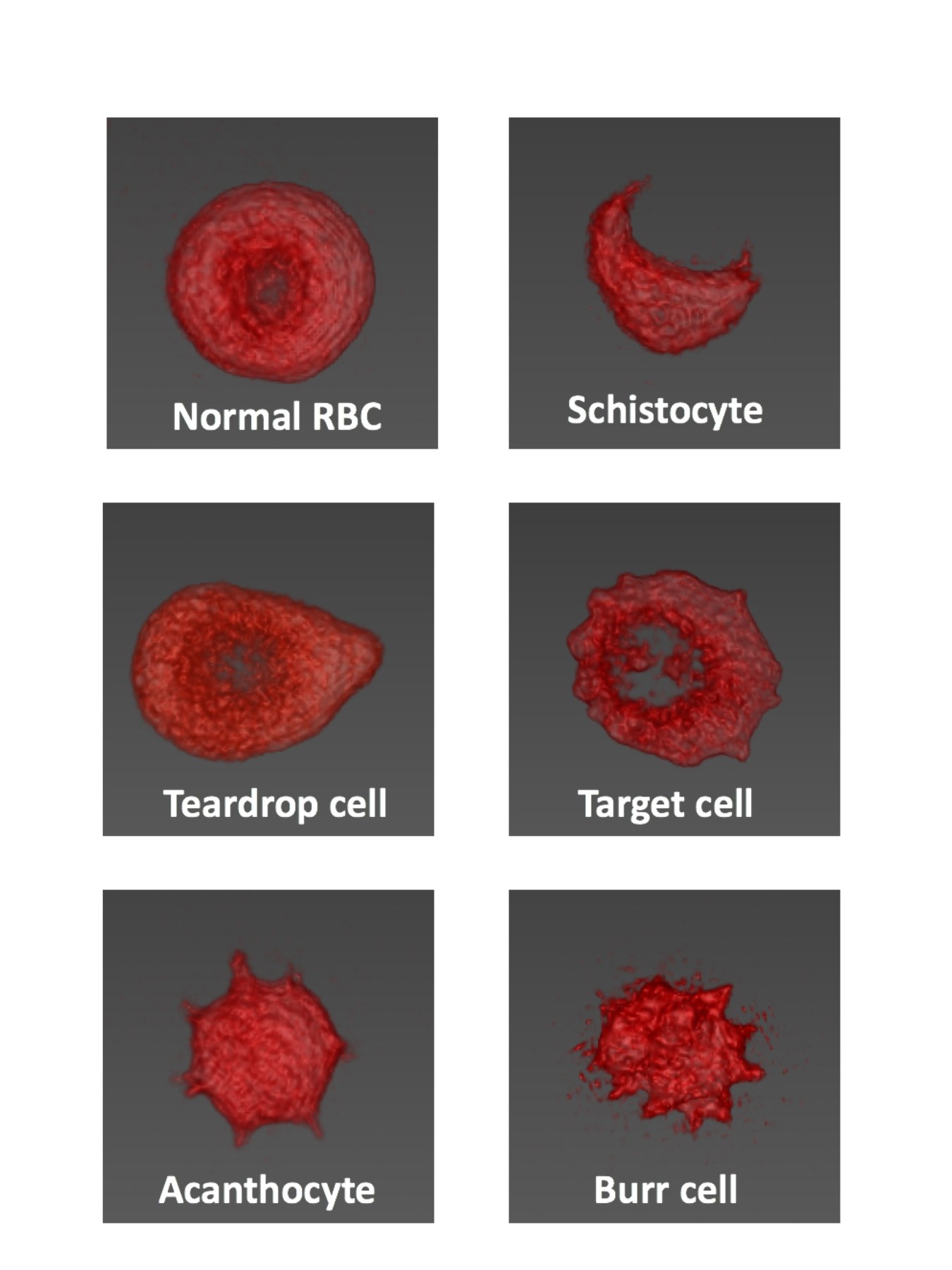
🔬 3D reconstruction of erythrocyte morphology using an optical diffraction tomography (ODT) microscope 📍 The study was conducted at Asan Medical Center and KAIST (South Korea)
🧪 Introduction: Scanning electron microscopy (SEM) is typically used to obtain 3D images of cells, but this is a labor-intensive process requiring special sample preparation. ODT is an interferometric microscopy technique that allows obtaining 3D tomograms of the refractive index (RI) of live cells without staining and fixation.
🧫 Methods: Blood remnants from EDTA tubes after complete blood count were used for analysis. Samples were diluted with DPBS buffer and applied between coverslips. 3D tomograms were captured using an HT-1S microscope (Tomocube Inc.), visualization via TomoStudio software.
🧍♂️ Results: 3D images of individual erythrocytes, including various forms of poikilocytes, were obtained. Cell morphology was clearly visualized at the single-cell level.
🧠 Conclusions: ODT is a fast and powerful tool for assessing erythrocyte morphology. It can be particularly useful in cases of questionable automated analyzer results, as well as for studying the pathogenesis of abnormal erythrocyte forms in live cells.
👉 Source: Koo S. et al. RECONSTRUCTED 3-D MORPHOLOGIES OF ERYTHROCYTES WITH OPTICAL DIFFRACTION TOMOGRAPHY SETUP MICROSCOPE //INTERNATIONAL JOURNAL OF LABORATORY HEMATOLOGY. – 111 RIVER ST, HOBOKEN 07030-5774, NJ USA : WILEY, 2018. – Т. 40. – С. 77-77. 🩸 The full article from the international journal of laboratory hematology is available in the library of blood and bone marrow mysteries.


