

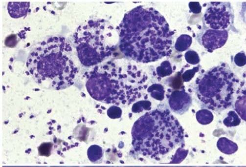
🔬 VETERINARY CYTOLOGY 🐶 Leishmania spp.: numerous amastigotes are detected within macrophages and freely in the smear background; they are oval, approximately 2–4 µm in size, with small purple nuclei, tiny rod-shaped kinetoplasts, and pale cytoplasm. Animal species: dog (a typical host for Leishmania spp.).
Source: Francesco Cian - Differential Diagnosis in Small Animal Cytology The Skin and Subcutis, 2018 Photo source: 🏛 Library “Veterinary Laboratory Medicine”


