

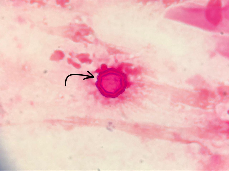
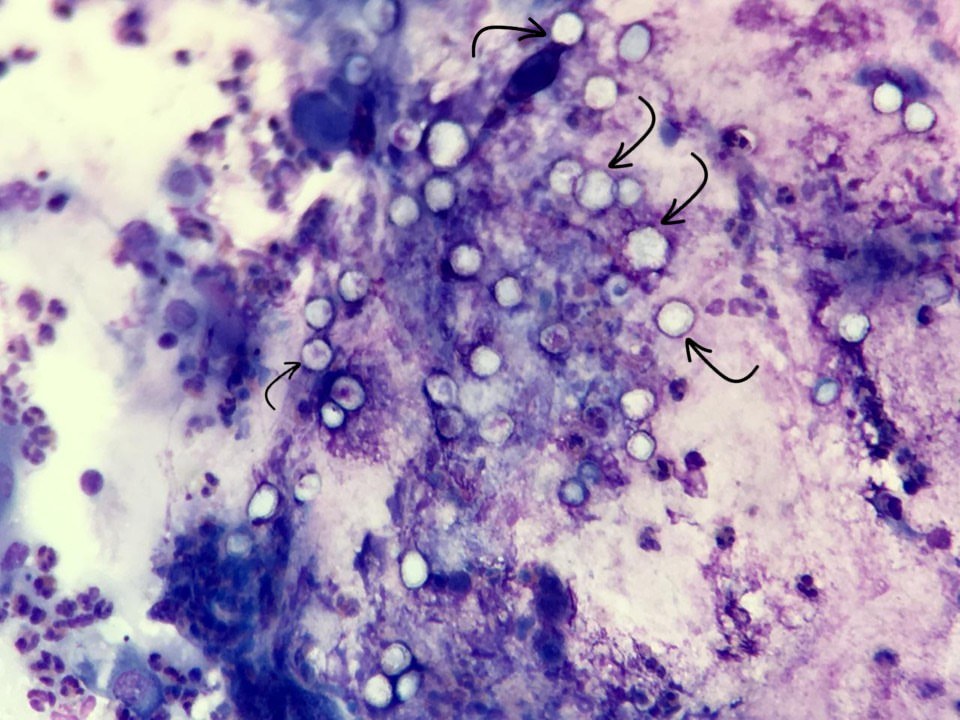
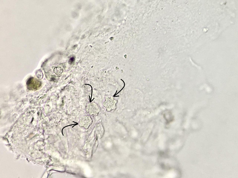
The images show Acanthamoeba cysts found in a corneal scrape from a lesion, confirming the diagnosis of amoebic keratitis. Acanthamoeba is a free-living protozoan widely distributed in water, soil, and air. Keratitis caused by this parasite is characterized by severe eye pain, redness, photophobia, tearing, and blurred vision.
The main risk factors include contact lens use (especially with inadequate hygiene), contact with contaminated water (swimming pools, tap water, showers, lakes, etc.), and eye trauma.
Acanthamoeba cysts are extremely resistant to disinfectants and can survive in poorly cleaned contact lens solutions. They are also resistant to many standard treatments, so early diagnosis is crucial for timely initiation of adequate therapy and prevention of potential vision loss.
The first preparation is Gram-stained, the second is Romanowsky-Giemsa stained, and the third is examined in a native preparation.


