

Amyloid bodies in prostatic secretion. Native preparation, 40x magnification + phone magnification
🙏 Photos kindly provided by Clinical Laboratory Diagnostics Doctor, Irina Selivanova LLC “Citilab-Ural”, Yekaterinburg
Microscopy of native prostatic secretion: morphology of amyloid bodies, mucus, and cellular elements at 40x magnification. A guide for laboratory hematology.
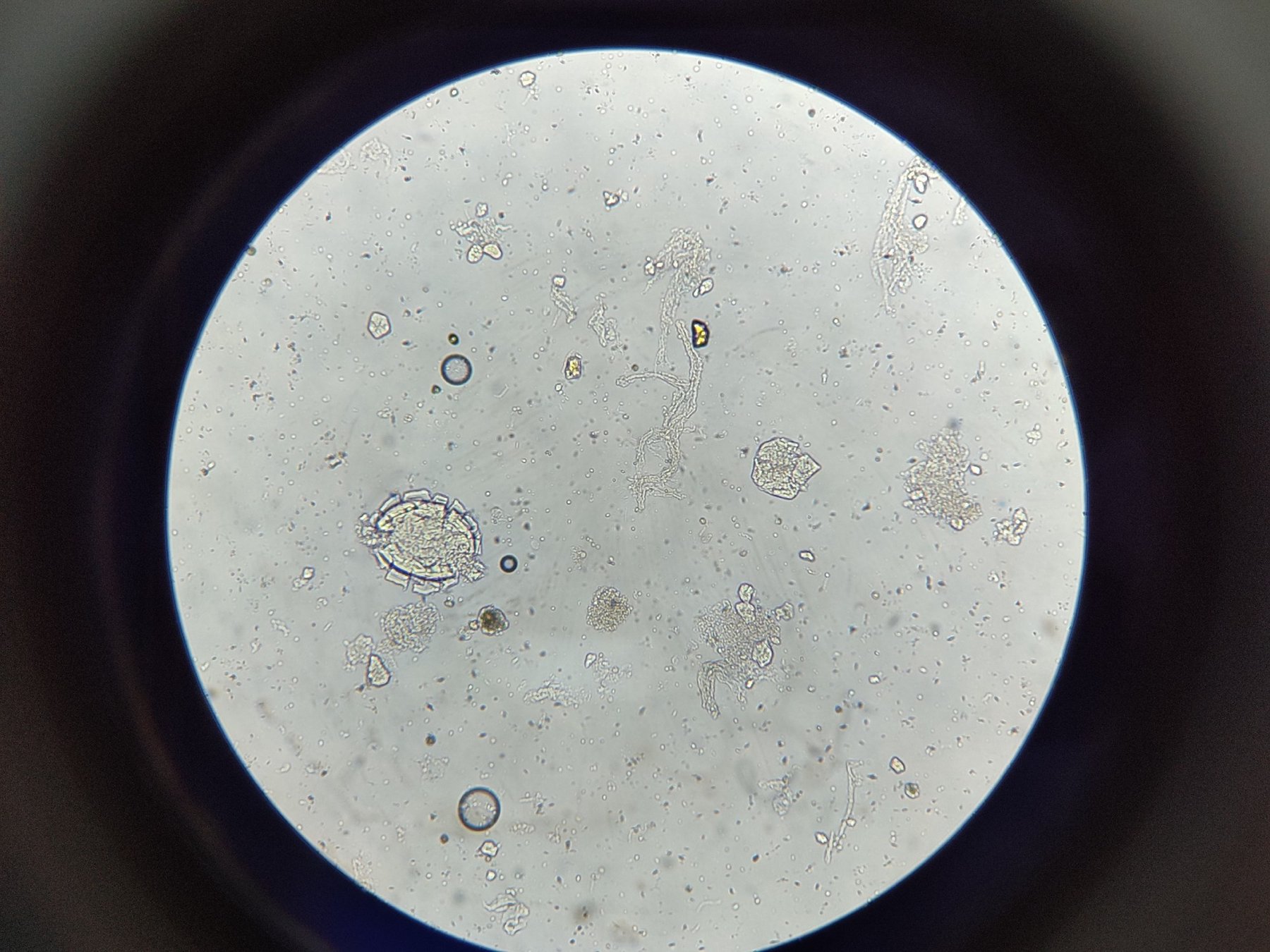
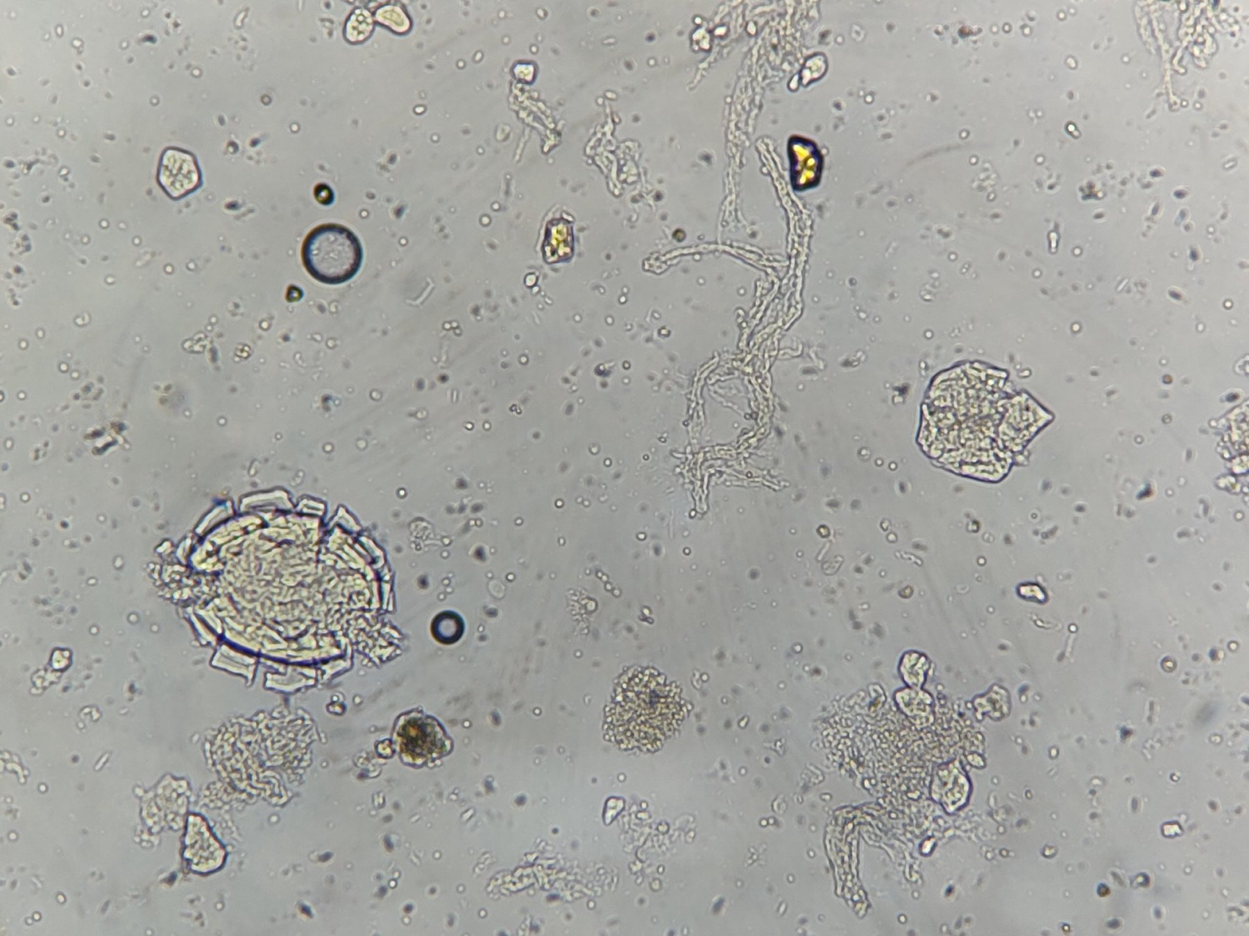
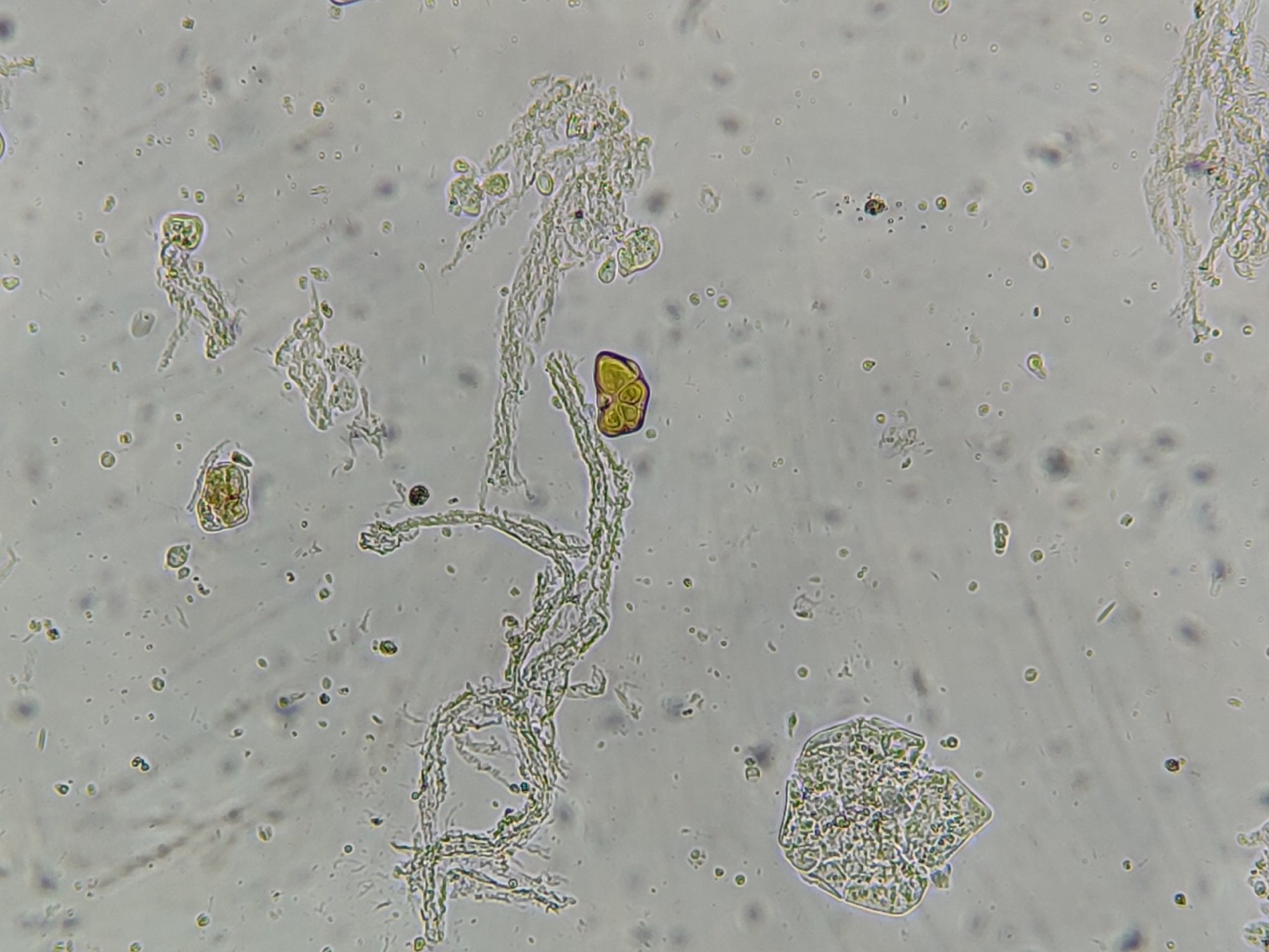
Amyloid bodies in prostatic secretion. Native preparation, 40x magnification + phone magnification
🙏 Photos kindly provided by Clinical Laboratory Diagnostics Doctor, Irina Selivanova LLC “Citilab-Ural”, Yekaterinburg

Morphology of amyloid bodies in native prostate gland secretion: concentric layering and clinical significance of the finding during microscopy.

Microscopy of the third portion of post-massage urine sediment: detection of prostatic amyloid bodies with characteristic layering during laboratory examination.

Morphology and microscopy of amyloid bodies in post-massage urine sediment. Features of detecting layered formations during prostate secretion microscopy.