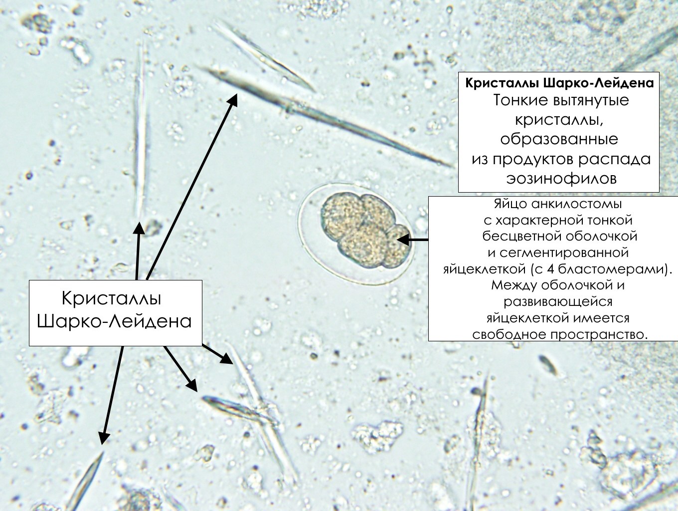
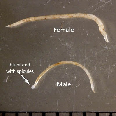
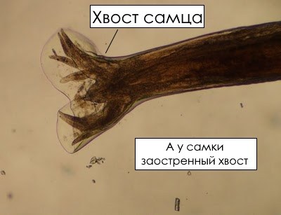
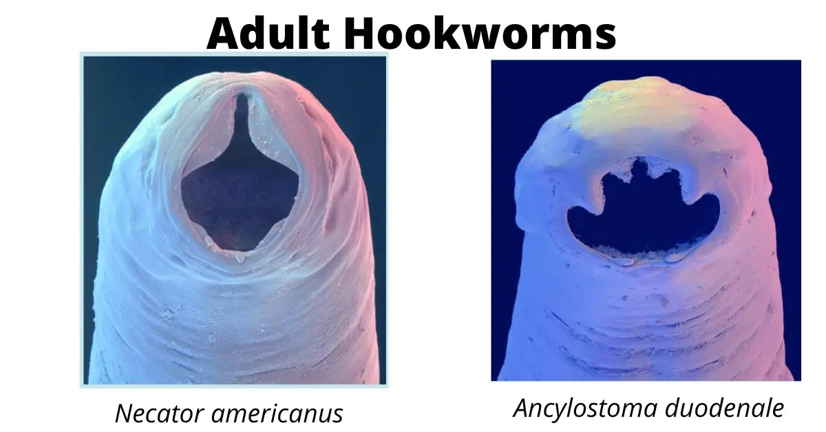
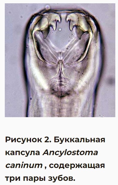
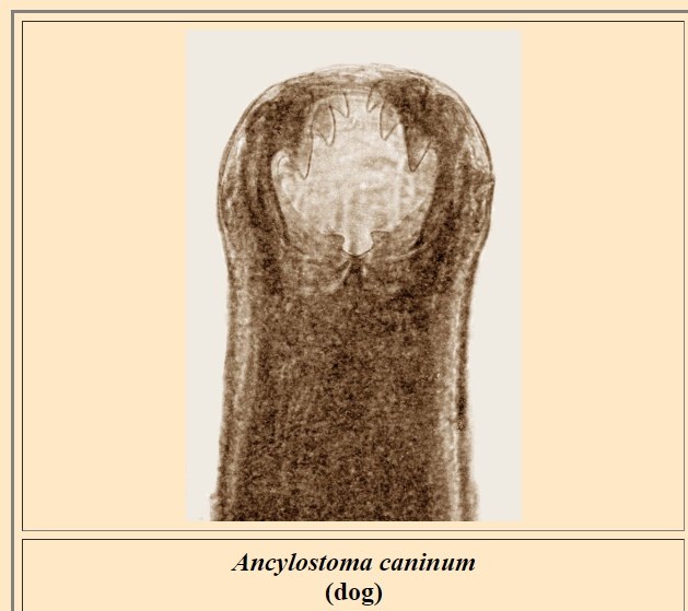
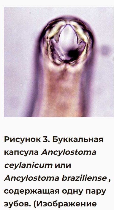
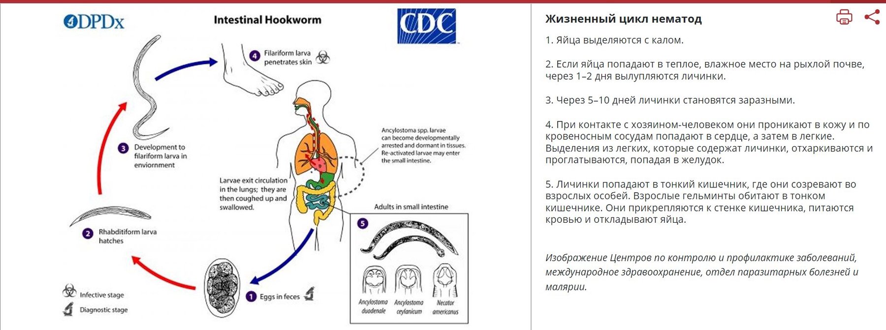
🐍Correct answer to the parasitology problem. ✅Hookworm: Ancylostoma duodenale. The most important clinical symptom is anemia, leading to high childhood morbidity in endemic areas. Severe infections can lead to impaired cognitive function and severe growth retardation. In pregnant women, severe infections can lead to prematurity and intrauterine growth retardation. ✅Differential diagnosis includes infection with the nematode Necator americanus and, less commonly, Ancylostoma caninum, the common dog hookworm. They can be distinguished by their oral apparatus: N. americanus has cutting plates instead of teeth, and A. caninum has 3 pairs of teeth instead of 2. Ancylostoma ceylanicum or Ancylostoma braziliense have 1 pair of teeth (photos 4-7). 👉HOW TO DISTINGUISH? ✅This is indeed Ancylostoma duodenale, which has a pair of two vampire teeth instead of a pair of three. Two ventral plates with two cusps each distinguish it from A. caninum, which has ventral plates with three cusps each. The larger worm is likely female, and the smaller is male. 1.3 cm is almost the upper size limit for adult females. 🔬 The image shows an egg with a very low number of blastomeres (only 4!). Such a low level of cleavage in feces is usually more characteristic of A. duodenale.
Ancylostoma duodenale: Morphology and Lab Diagnosis
Identification criteria for Ancylostoma duodenale: egg morphology, Charcot-Leyden crystals in stool, oral capsule structure, and differentiation from Necator americanus.