

Sand from under the free edge of the toenail plate. Beautiful, but it makes viewing the sample difficult.
🙏 Material kindly provided by medical laboratory technician Dmitry Emelyanov.
Sand particles in a nail plate scraping sample: a common artifact in onychomycosis microscopy, hindering visualization of fungal mycelium and spores.
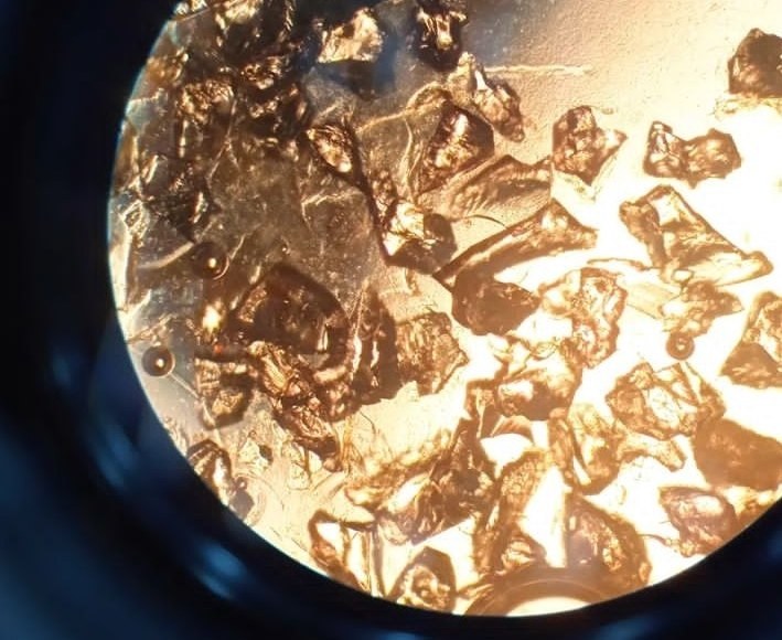
Sand from under the free edge of the toenail plate. Beautiful, but it makes viewing the sample difficult.
🙏 Material kindly provided by medical laboratory technician Dmitry Emelyanov.

Microscopy of nail scrapings with 20% KOH: why myelin is mistaken for fungal spores, morphological differences of artifacts, and sample preparation features.

Laboratory detection of Candida fungi: microscopy of scrapes with 20% KOH. Blastospores, pseudomycelium of the fungus in onychomycosis and candidal paronychia.

Microscopy of skin scrapings in 20% potassium hydroxide (KOH) solution: erythrocyte changes and features of differential diagnosis of artifacts.