

🔬 Ascitic fluid. Romanowsky stain. Atypical cells in large numbers. Woman, ovarian C-r.
🙏 Photos kindly provided by KDL biologist Yulia Guseva GBUZ Belorechenskaya CRB.
Cytological findings in ascitic fluid in ovarian cancer. Romanowsky stain: clusters of atypical cells, cellular polymorphism, and vacuolization.
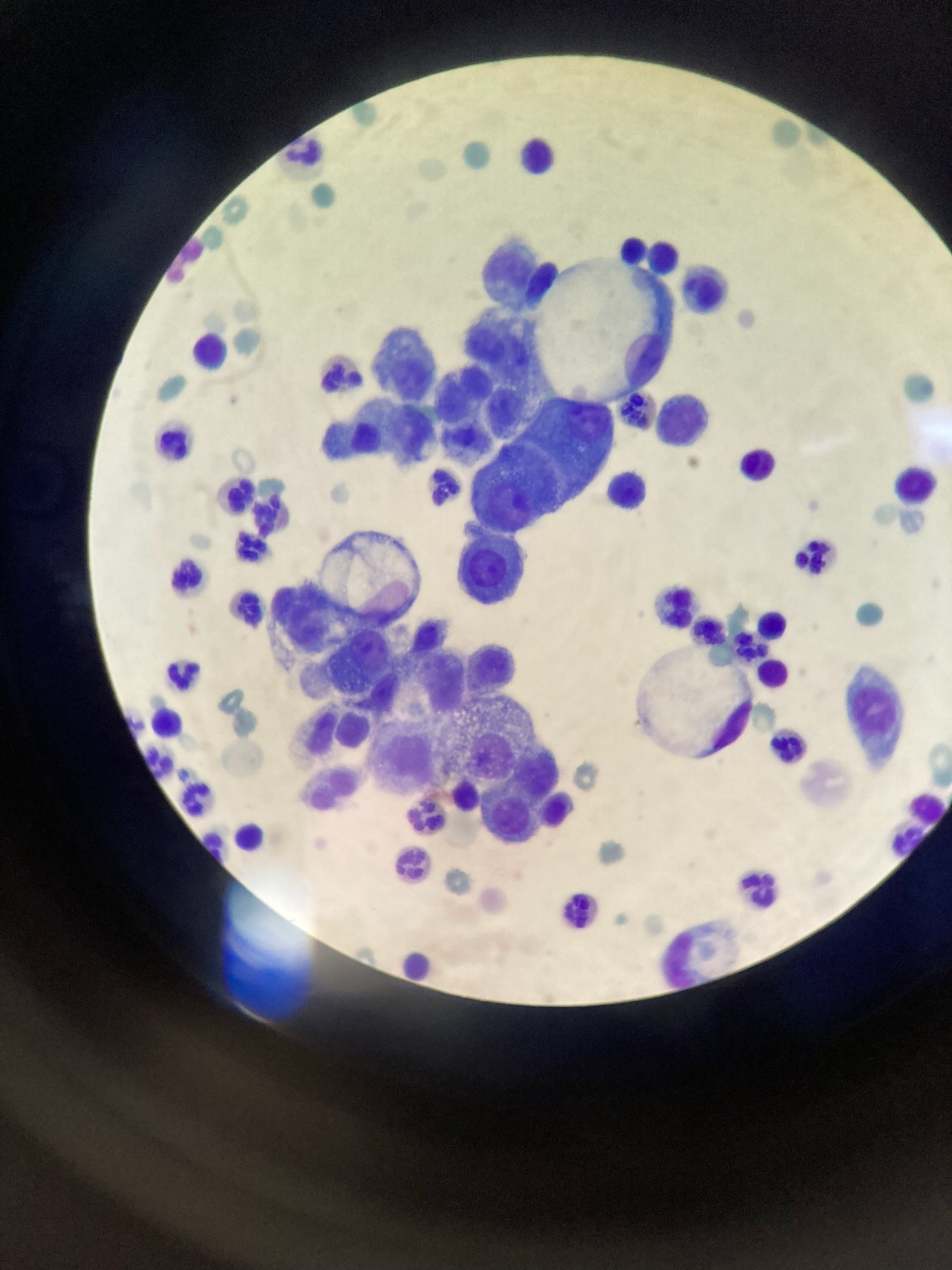
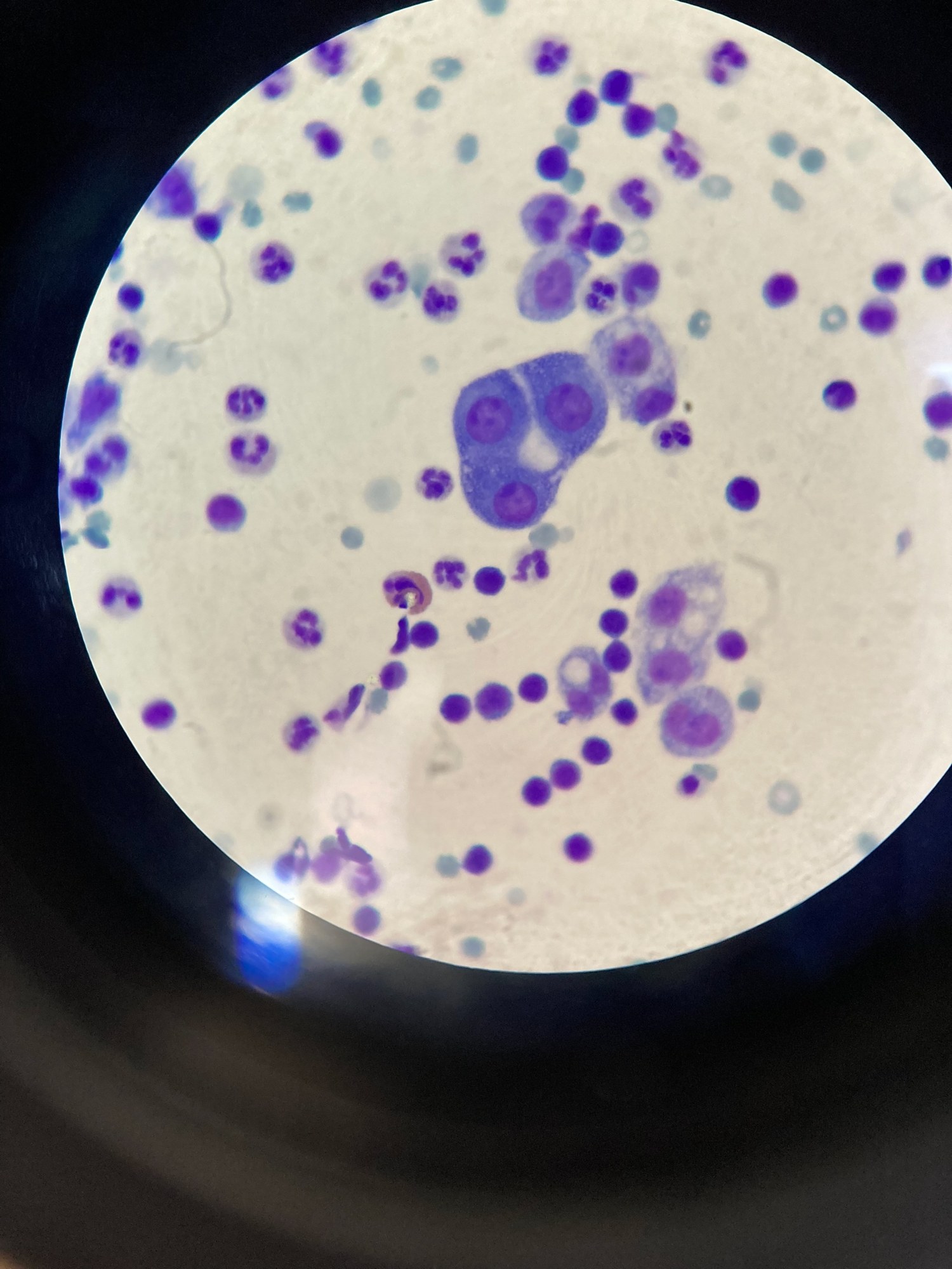
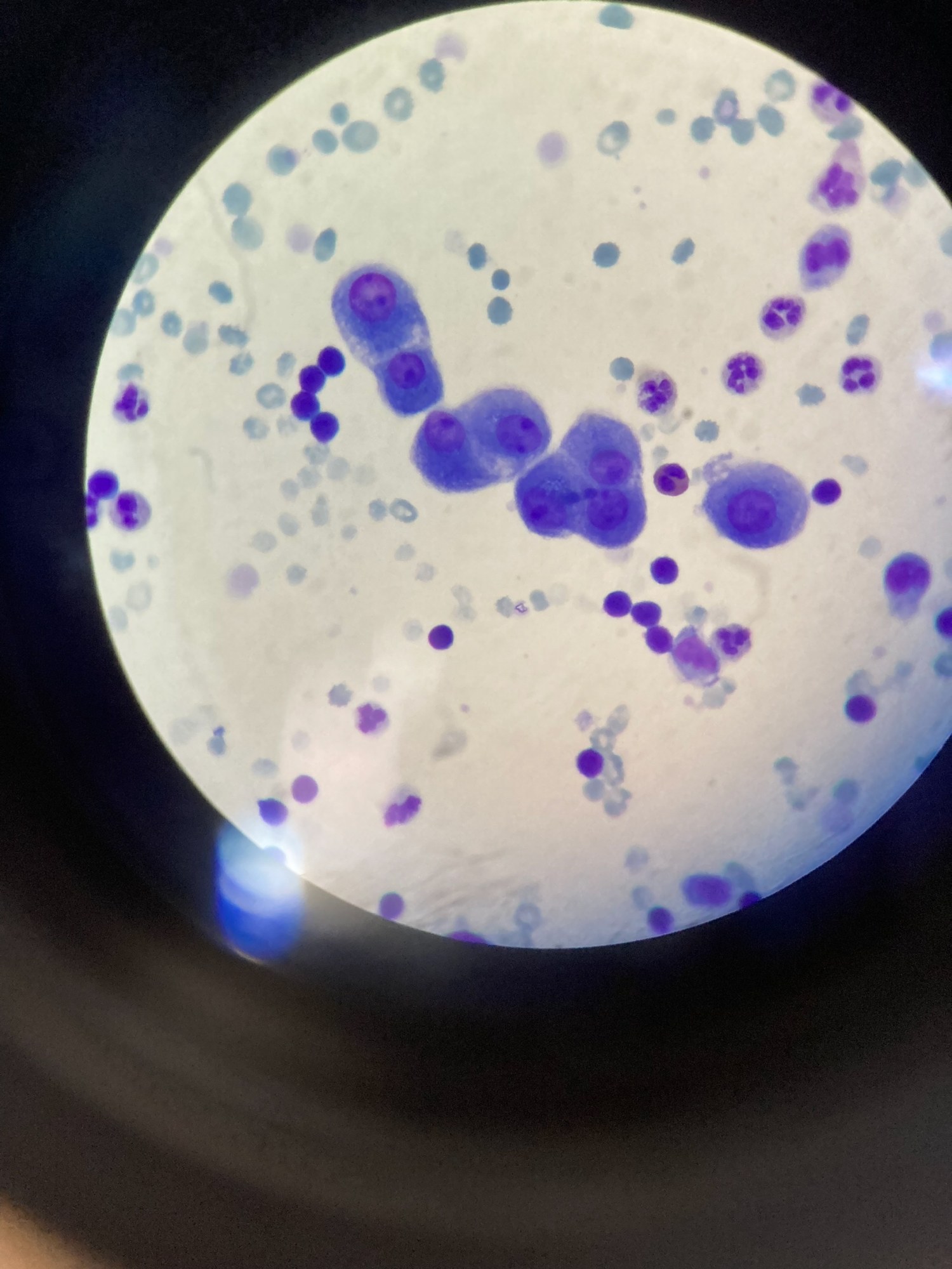
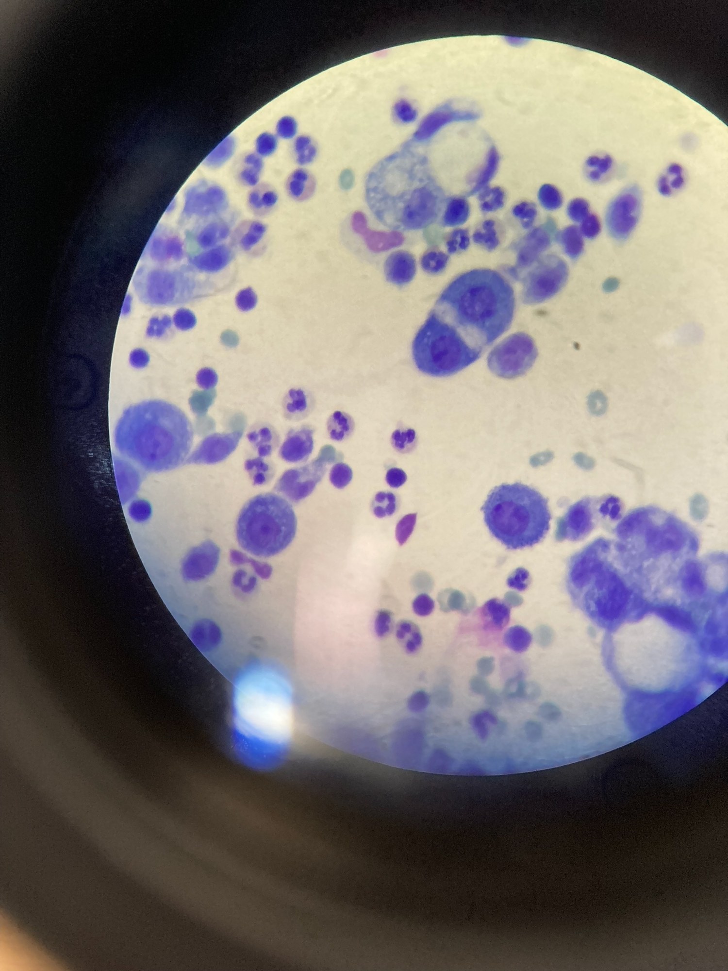
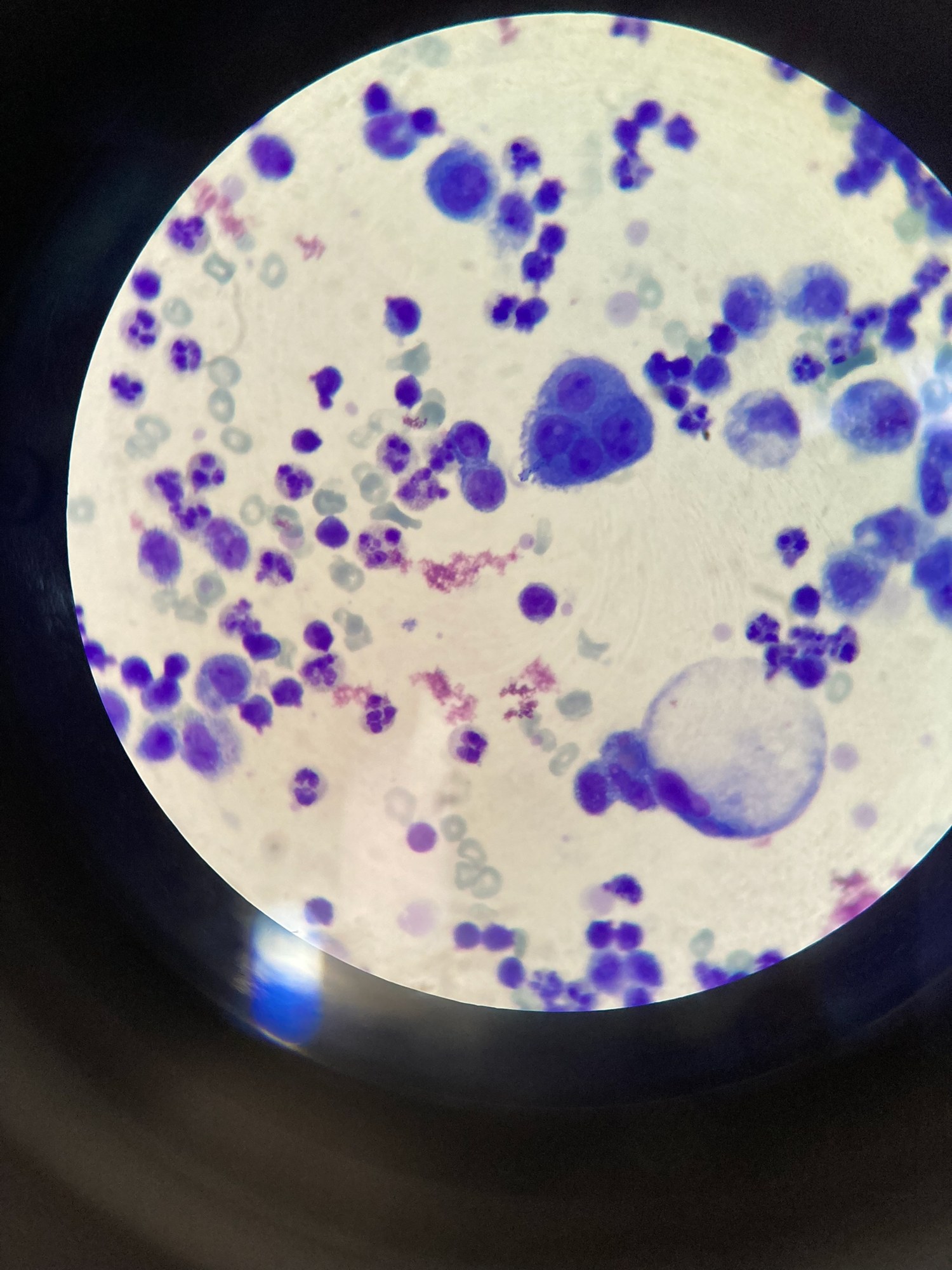
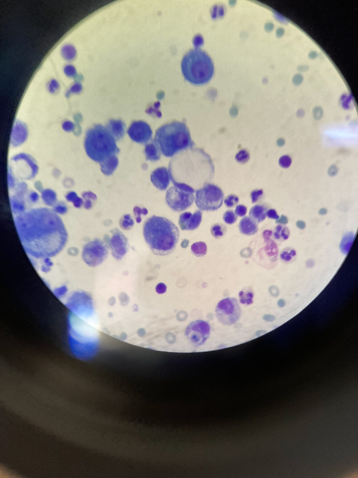
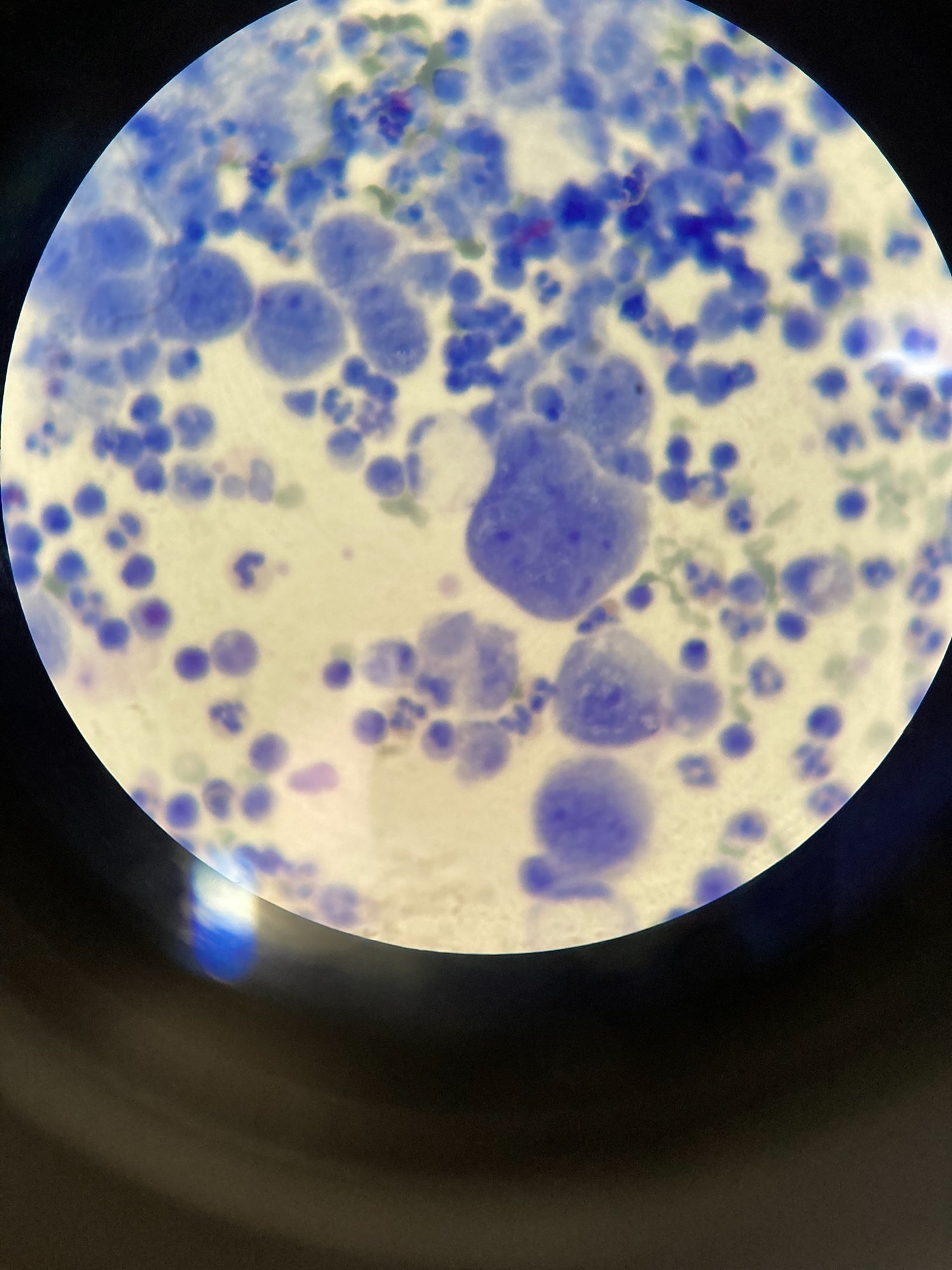
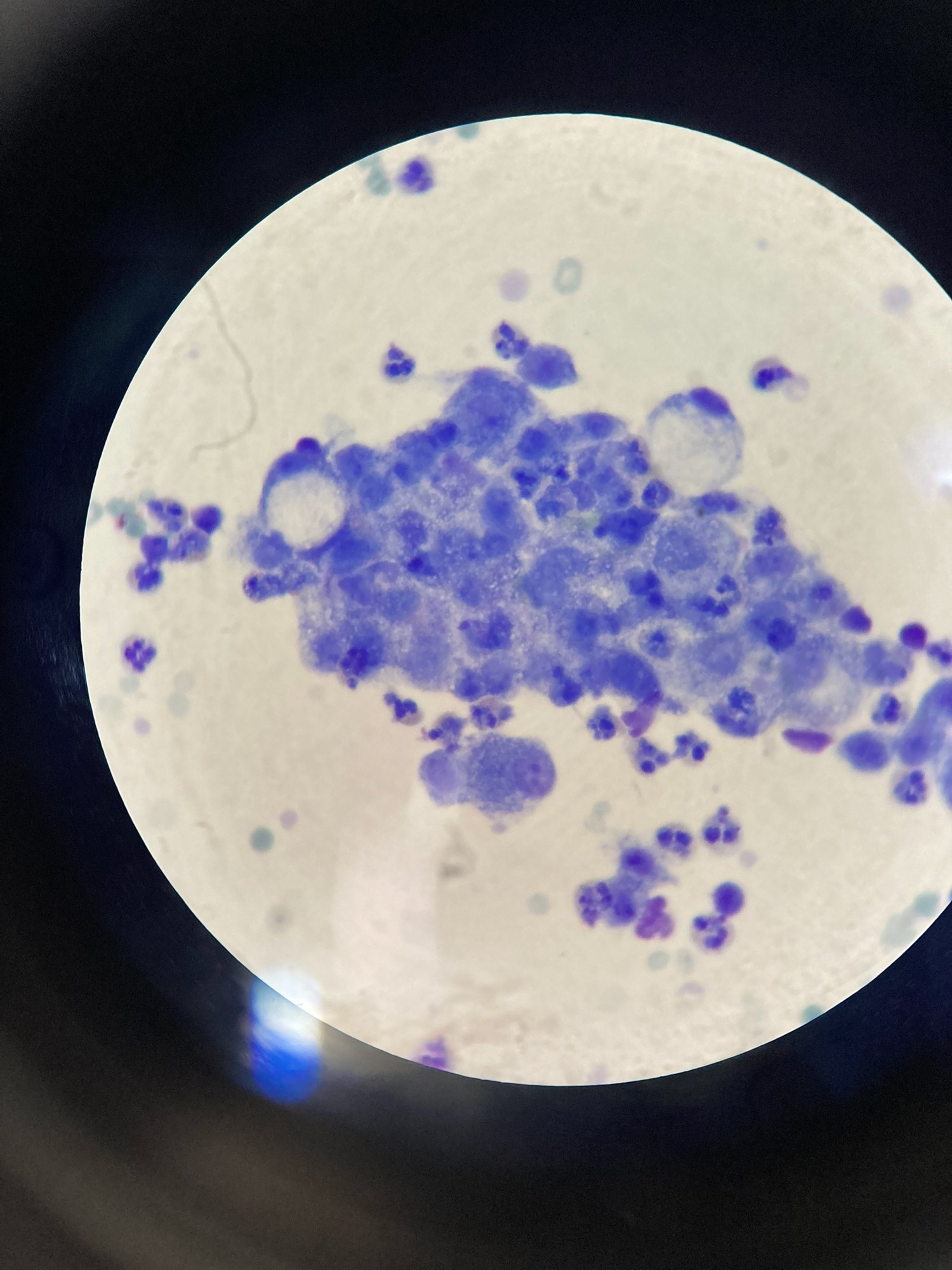
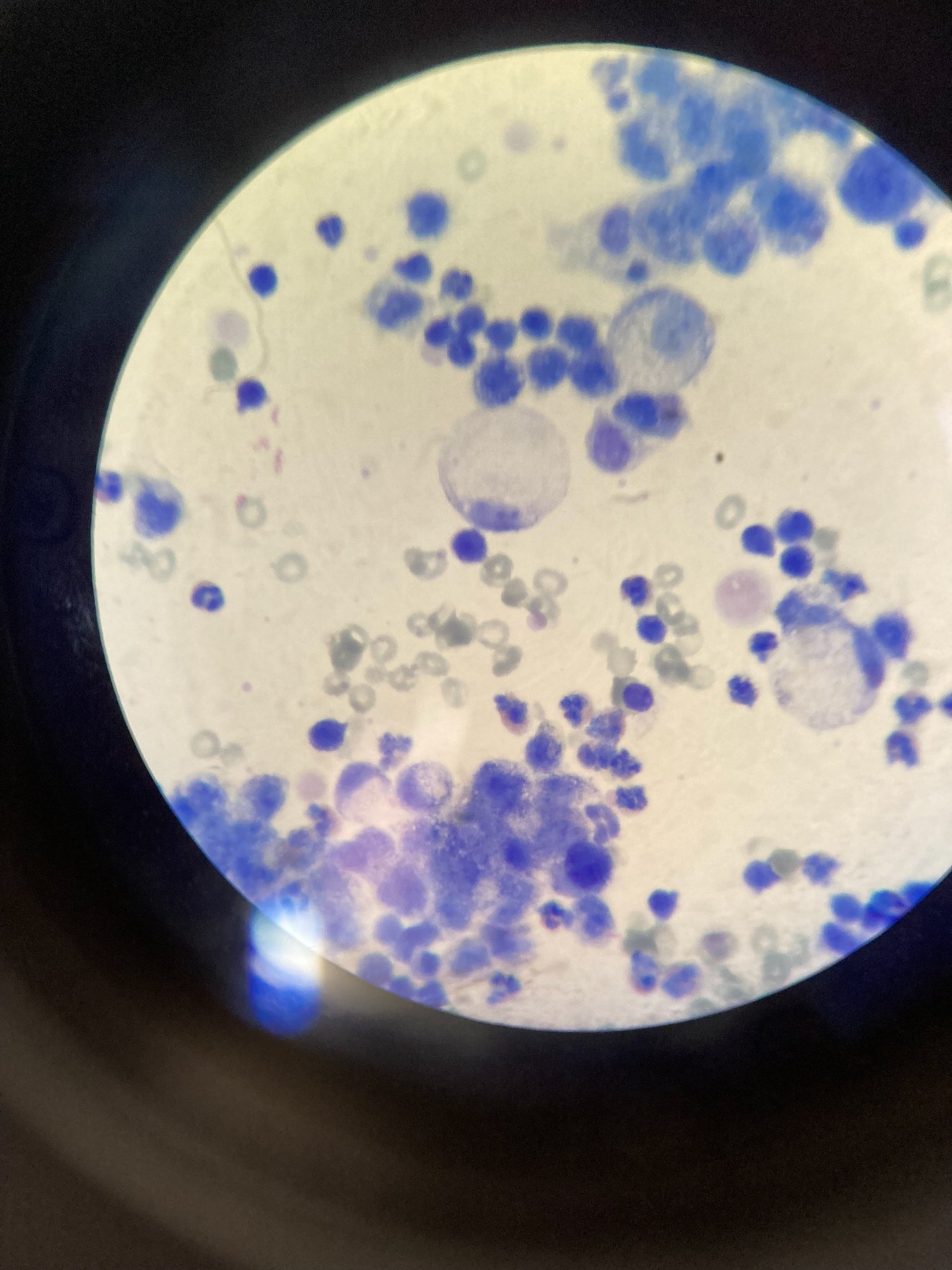
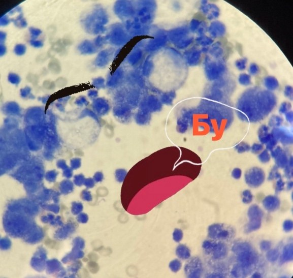
🔬 Ascitic fluid. Romanowsky stain. Atypical cells in large numbers. Woman, ovarian C-r.
🙏 Photos kindly provided by KDL biologist Yulia Guseva GBUZ Belorechenskaya CRB.

Cytological examination of ascitic fluid in ovarian cancer: microscopy of native preparations and smears stained with Romanowsky-Giemsa.

Cytological examination of ascitic fluid: detection of ovarian adenocarcinoma cell complexes, malignancy criteria, and a clinical case study.

Microscopy of ascitic fluid sediment from a patient with ovarian cancer: cytological signs of metastatic involvement and atypical cell complexes.