
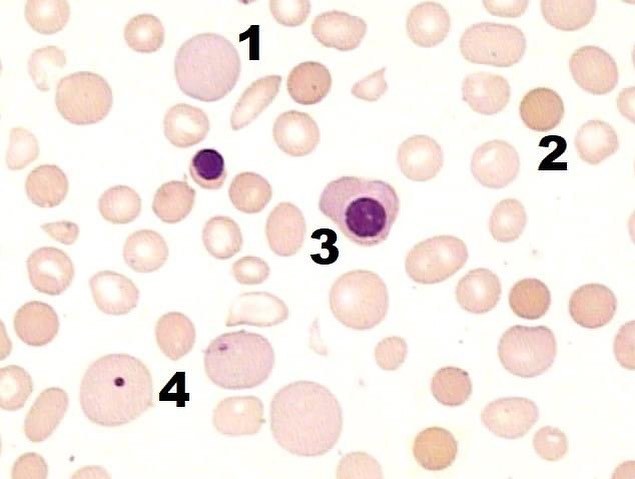
Autoimmune hemolytic anemia caused by warm antibodies
Polychromasia and spherocytes are found in the peripheral blood; circulating erythroblasts and occasional Howell-Jolly bodies may be present.
The disease is caused by immunoglobulin G class antibodies that react with red blood cells at temperatures of 37 degrees Celsius and above. This is the most common form of autoimmune hemolytic anemia.
It occurs in patients of all age groups and both sexes, but is more common in adult women, and is accompanied by anemia of varying severity and an enlarged spleen.
The direct antiglobulin test is positive, confirms the diagnosis, and may indicate the cause. The indirect antiglobulin test is negative.
Biochemical studies may show an increase in indirect bilirubin and lactate dehydrogenase activity, as well as a decrease in haptoglobin concentration.


