

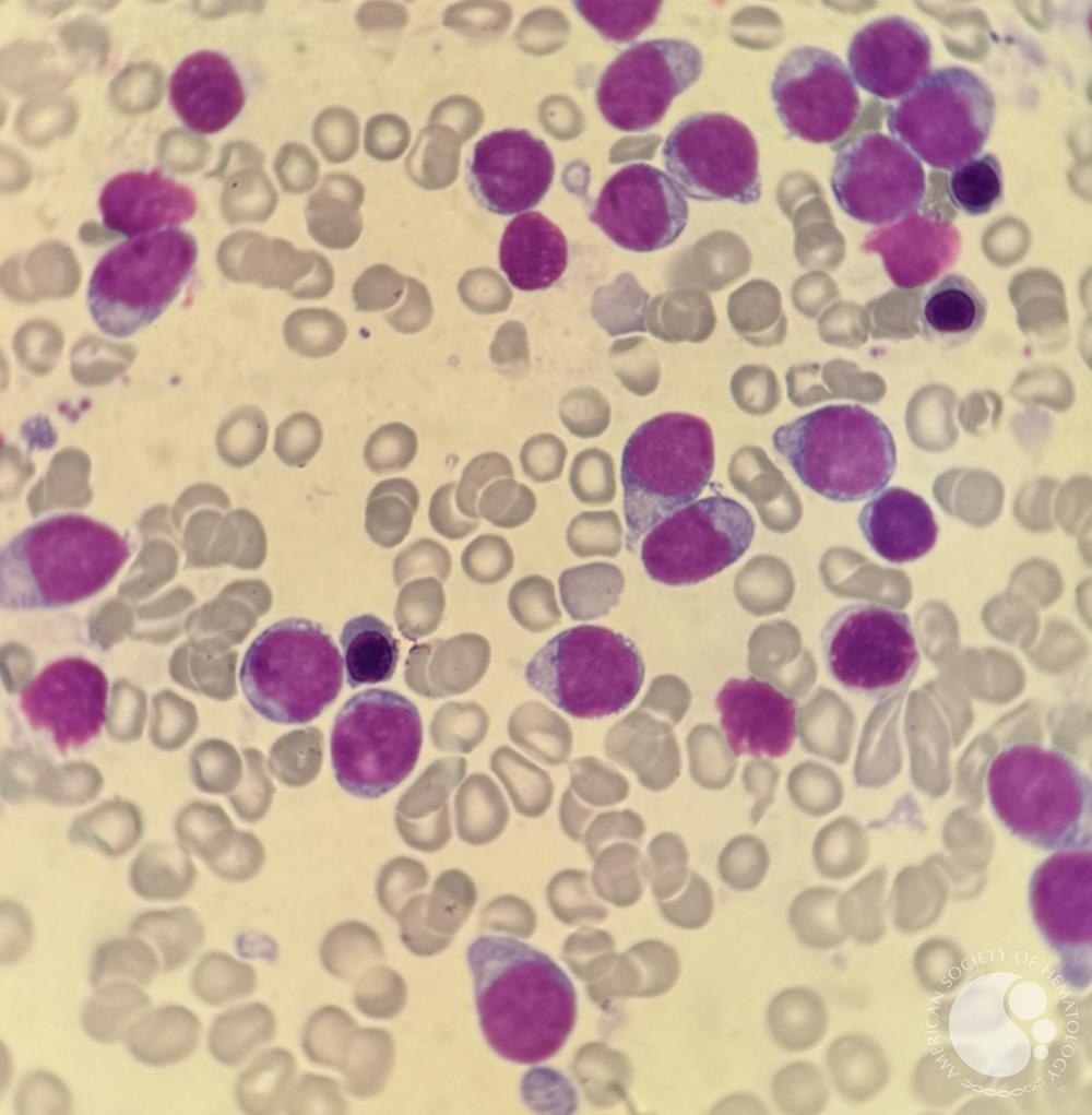
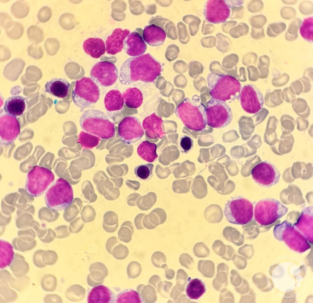
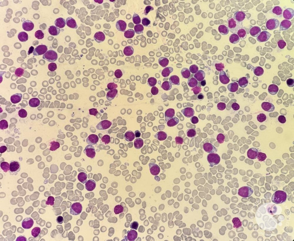
❗ Clinical case. Blastic Plasmacytoid Dendritic Cell Neoplasm (BPDCN) ✅ A 57-year-old woman presented with spontaneous bruising, some skin lesions and mild itching, and cytopenia of unknown cause. Numerous medium-sized blastoid cells were found in peripheral blood and bone marrow.
💡 Immunophenotyping of peripheral blood and bone marrow revealed an abnormal population with moderate CD45 expression and low side scatter, which showed positive expression for: CD56, CD7, CD33, HLADR, CD4, CD26, and NG2. And negative expression for CD34, CD19, CD10, CD5, CD2, CD3, CD25, CD30, CD16, CD57, TCRa/b, TCRg/d, CD1a, CD38, cytMPO, cytTDT, cytCD3, cytCD79a. This was reported as acute leukemia with no detected lineage-defining markers, suspected T-ALL given morphology and some T-cell markers.
✅ Further immunophenotyping later demonstrated CD123 positivity. This phenotype (CD7+ CD4+ CD56+ CD123+ HLADR+) is most characteristic of BPDCN, which is retrospectively confirmed by morphology and clinical picture.
✅ Blastic plasmacytoid dendritic cell neoplasm with peripheral blood involvement is an aggressive tumor originating from plasmacytoid dendritic cells with a high frequency of skin and bone marrow involvement and leukemic dissemination. Previously, BPDCN was called blastic NK-cell lymphoma/agranular CD4+ NK-leukemia.
👉 Source: imagebank.hematology


