

🔬 A solitary blast found in a peripheral blood smear.
🙏 Image kindly provided by Clinical Laboratory Diagnostics Physician, Anna Ivanova, Moscow.
Features of blast cell detection and morphology in peripheral blood smears: differential diagnosis during leukocyte formula microscopy.
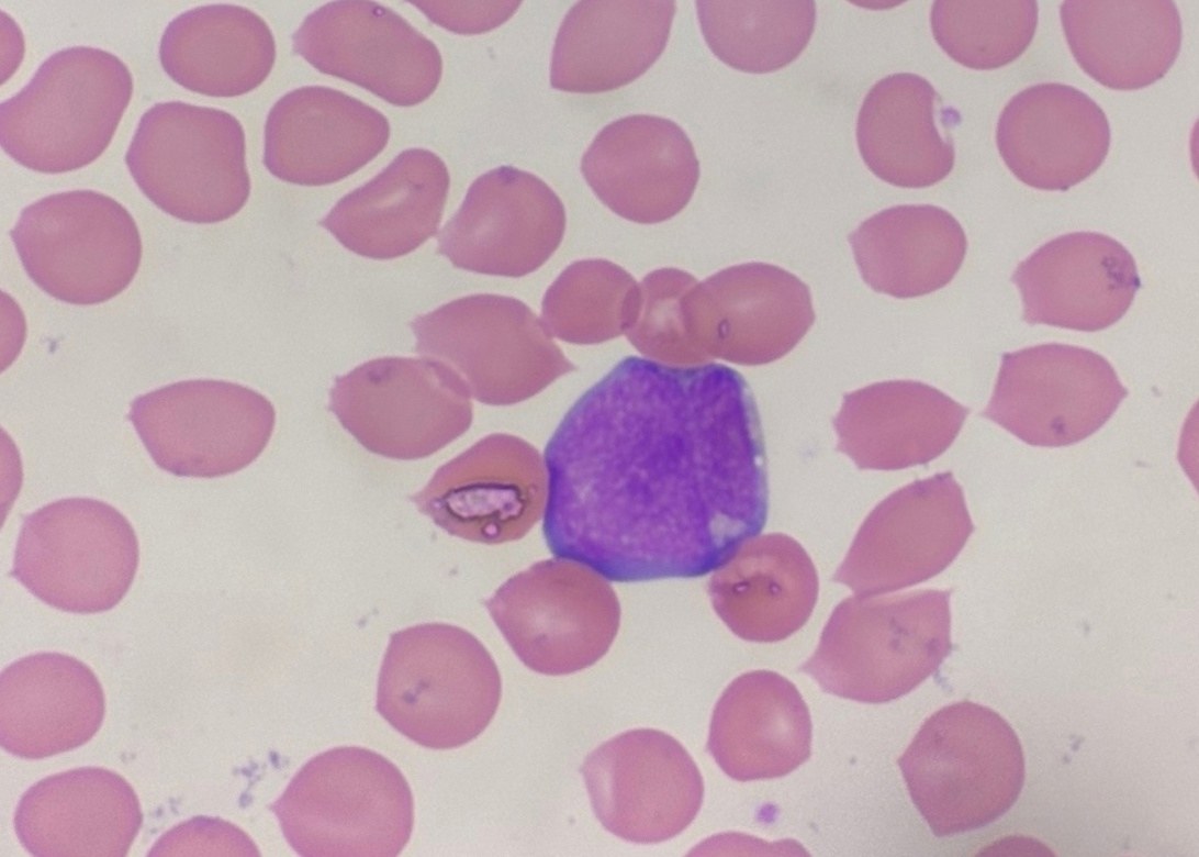
🔬 A solitary blast found in a peripheral blood smear.
🙏 Image kindly provided by Clinical Laboratory Diagnostics Physician, Anna Ivanova, Moscow.

Explore the morphology and arrangement of myeloblasts in a blood smear from a patient with acute myeloid leukemia without maturation, including microscopy and cytomorphology.

Blood smear in acute myeloblastic leukemia without maturation (FAB M1): pronounced leukocytosis, myeloblast morphology, and laboratory diagnostic criteria.

A case of acute leukemia in a rat: leukocytosis 258×10⁹/L, total infiltration with blast cells. Blood smear microscopy, Romanowsky-Giemsa stain.