

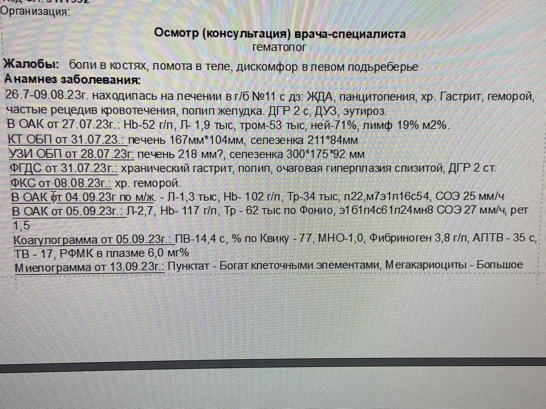
❗Clinical case of Gaucher Disease. An accidental finding! A lysosomal storage disease. ✅ A 51-year-old female patient was referred by a therapist to a hematologist with a diagnosis of iron deficiency anemia (? CBC in photo № 6). Her medical history included frequent recurrent bleeding and a gastric polyp. She complained of bone pain, body aches, and discomfort in the left hypochondrium.
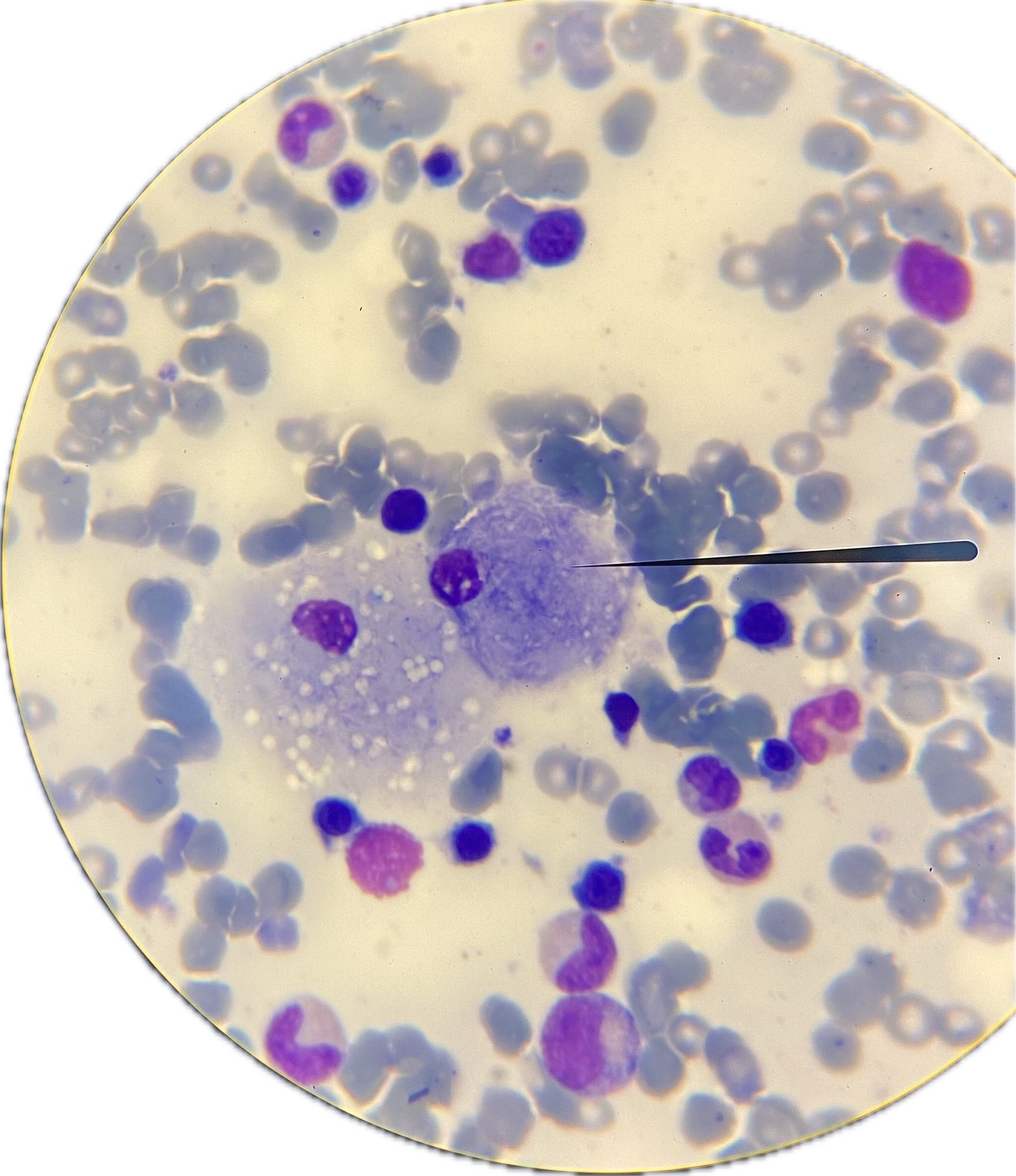
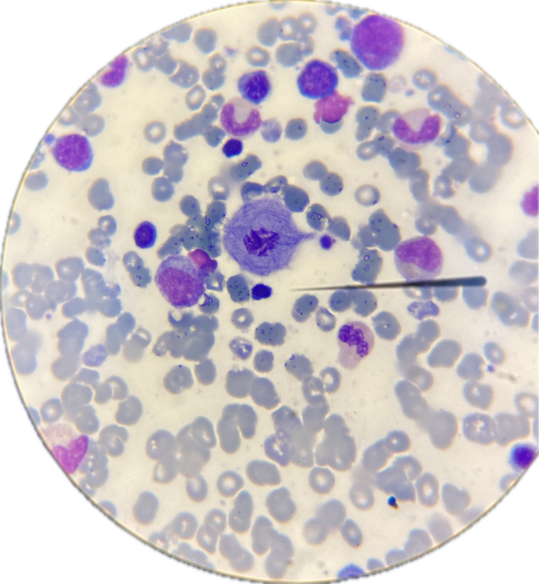
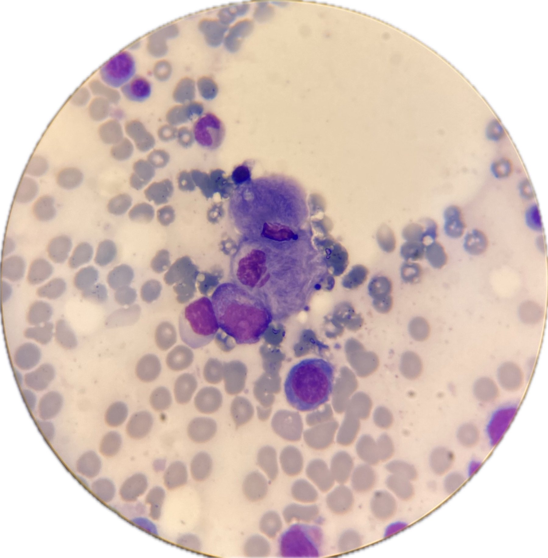
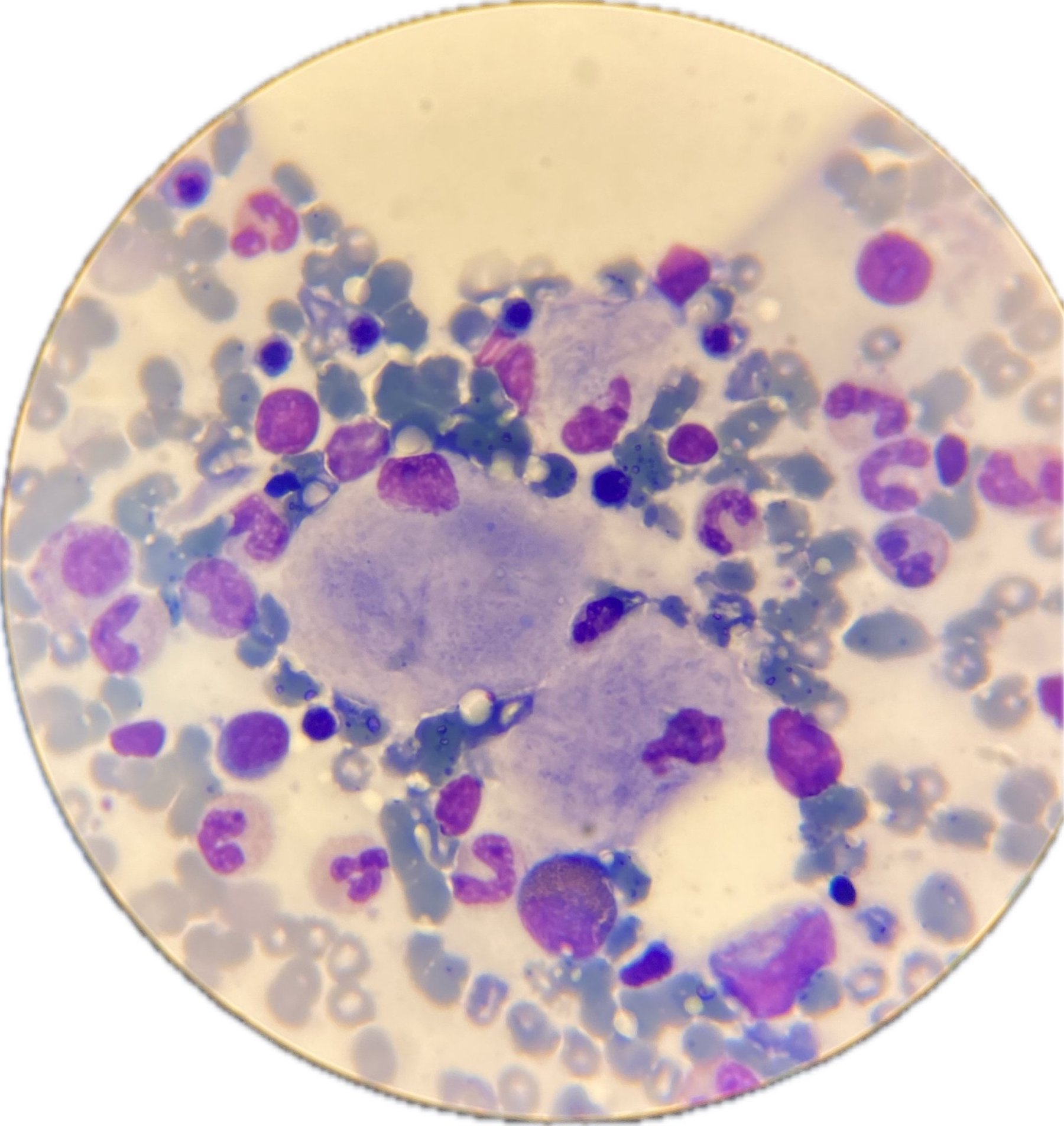
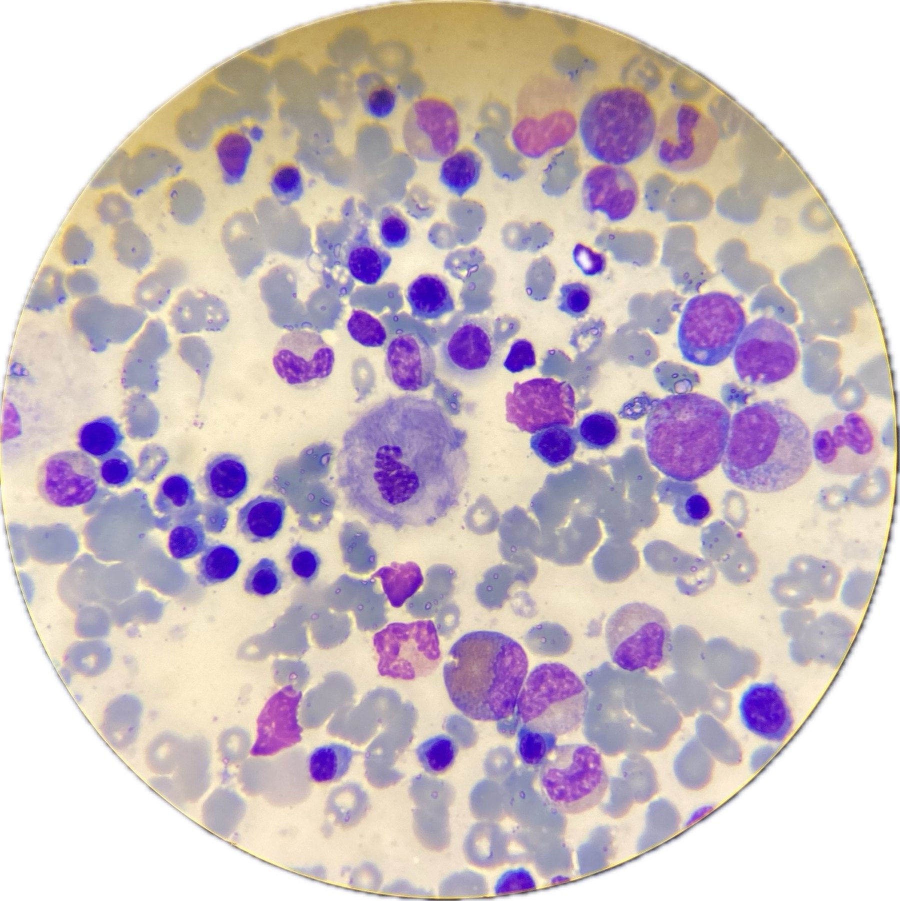
🦴Myelogram Bone marrow is hypercellular, with a large number of megakaryocytes, expanded erythroid lineage, and normoblastic hematopoiesis. Signs of dysplasia in the erythroid lineage – nuclear abnormalities, multinuclearity – 5%. The bone marrow smear shows a large number of “Gaucher-like” macrophages, lipophages (photos № 1-5).
✅Selective screening for storage diseases was performed: The study results revealed an increased hexosylsphingosine concentration – 525 ng/ml (0.2-1.0), and decreased glucocerebrosidase activity – 0.49 µmol/h (1.5-25). Chito-triosidase activity was sharply elevated – 4043 nM/ml/h (2.5-100).
✅ A full analysis of the GBA gene was performed: autosomal recessive inheritance. A nucleotide sequence variant c.1226A>G was found in exon 10 of the gene in a heterozygous state, described in the HGMD database as pathogenic. A nucleotide sequence variant c.1448T>C was found in exon 11 of the gene in a heterozygous state, described in the database. These changes are characteristic of Gaucher disease. The diagnosis was confirmed.
✅ Consulted by phone with the head of the Department of Orphan Diseases of the National Medical Research Center for Hematology, Moscow; hospitalization to the NMRC was recommended.
👉The clinical case was kindly provided by Marina Boyarskaya, a laboratory diagnostic doctor at the Regional Clinical Hospital, Barnaul.


