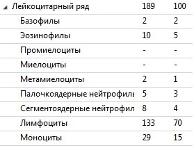
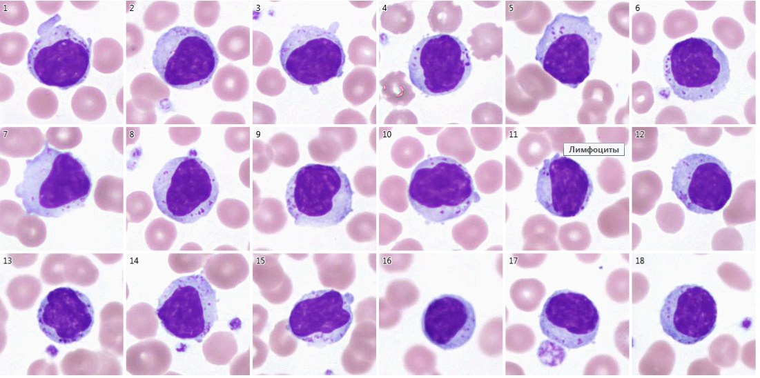
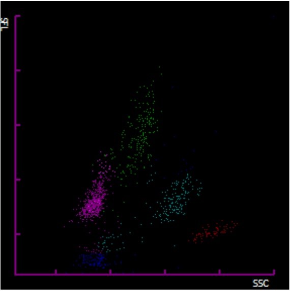
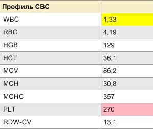
LEUKOPENIA WITH A RELATIVE INCREASE IN LARGE GRANULAR LYMPHOCYTES. Differential leukocyte count, or leukocyte formula, is the percentage of all leukocyte classes in the blood. In leukopenia, it is usually very difficult to count the differential leukocyte count using a conventional microscope, because the fewer leukocytes in the smear, the harder it is to find these 100 cells. However, counting the differential leukocyte count in this case is very necessary to understand which fraction of neutrophils or lymphocytes caused the decrease in the number of leukocytes. Also, as we know, hairy cell leukemia occurs more often in leukopenia. Therefore, the detection of “hairy” lymphocytes or blasts will also help to understand the true cause of the decrease in leukocytes and to timely prescribe bone marrow aspiration or flow cytometry, if necessary. A CASE FROM MY PRACTICE. A blood sample for a complete blood count from a 72-year-old female patient was delivered to the laboratory, WBC = 1.33×10^9/L. The WDF scattergram showed an increase in the “tail” in the monocyte region. A decision was made to perform microscopy. The Sysmex SP-1000 automatic slide stainer prepares 2 blood smears in cases of leukopenia, which were loaded into the Hema Vision automatic microscopy system. The program combines photos of 2 smears under one barcode into 1 differential leukocyte count, which is very convenient. In addition to a relative increase in lymphocytes and severe absolute neutropenia, an increase in the number of large granular lymphocytes was detected (Viral infection? T-cell large granular lymphocytic leukemia? Chemotherapy?). As we know, large granular lymphocytes (LGLs) help the body fight viruses and also tumor cells. The laboratory did not specify the diagnosis for this patient. The result was issued with a note on the number of LGLs – 18%.
Large Granular Lymphocytes in Leukopenia Blood Smear
Features of differential leukocyte count in severe leukopenia: detection of large granular lymphocytes, WDF scattergram analysis, and digital microscopy.