

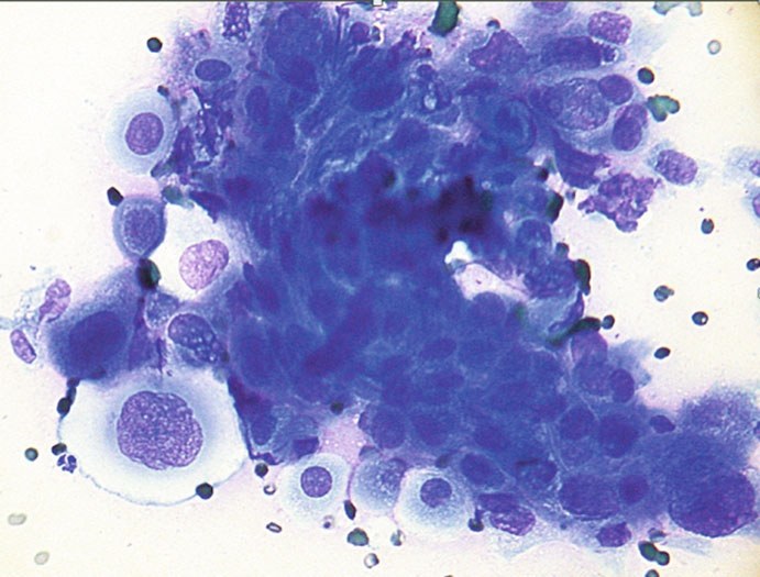
🔬 VETERINARY CYTOLOGY SKIN METASTASES. Groups of tumor bronchial cells with pronounced cellular atypia and signs of squamous differentiation.
Skin metastases of internal organ tumors (not primary skin tumors) in animals. Such metastases are rare and are mainly described in carcinomas of the stomach, intestines, prostate, bronchi, testicular seminoma, and some sarcomas (hemangiosarcoma, osteosarcoma). The spread of tumor cells occurs predominantly hematogenously, so lesions can appear in any area of the skin.
The most well-known example is bronchogenic carcinoma in cats, causing “lung–digit syndrome” with multiple metastases, most often in the digits and muscles.
Mammary gland tumors are usually considered separately; however, inflammatory mammary carcinoma is included in this chapter due to its cutaneous manifestations and lymphatic spread.
Round cell tumors (mastocytoma, lymphoma, histiocytic diseases, multiple myeloma) are not discussed, as skin and visceral lesions often coexist, making it difficult to determine the primary tumor; they were covered in the previous chapter. Photo source: 🏛 Library “Veterinary Laboratory Medicine”


