

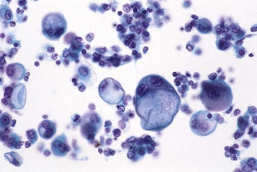
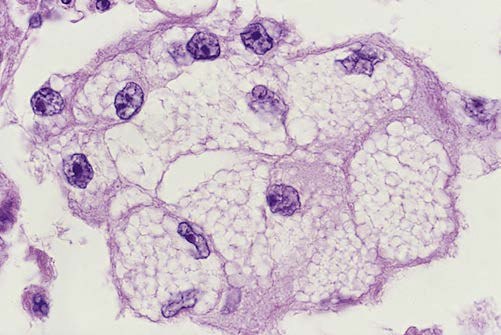
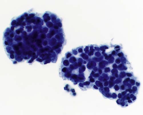
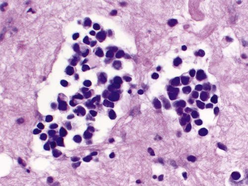
Cytology. Malignant Mesothelioma Cells. 🔬 Images 1 and 2: Malignant mesothelioma, unusual variants. In rare cases, mesotheliomas show marked cytoplasmic vacuolization (“clear cell type”). 🔬 Images 3 and 4: Some mesotheliomas consist of small cells; however, the use of the term “small cell mesothelioma” is not recommended to avoid confusion with small cell lung carcinoma.
(1 and 3 – Papanicolaou stain; 2 and 4 – hematoxylin and eosin stain).
WHERE ARE THESE CELLS FOUND?
- Pleural effusion – the most common source. Mesothelioma is often accompanied by fluid accumulation in the pleural cavity.
- Ascites (peritoneal effusion) – in peritoneal mesothelioma.
- Pericardial effusion – in pericardial mesothelioma (rare).
Mesothelium is an epithelial tissue lining the serous membranes of body cavities (peritoneum, pleura, pericardium) in vertebrate animals and humans. It develops from the mesoderm.
💎 Find even more useful cytology materials in our LAB-TESTS library. A link with access to the library is automatically sent to you via private message after paying for a VK Donut Labformula subscription.
Source of images for publication: Cibas - Cytology. Diagnostic Principles and Clinical Correlates 5 ed (2021)


