

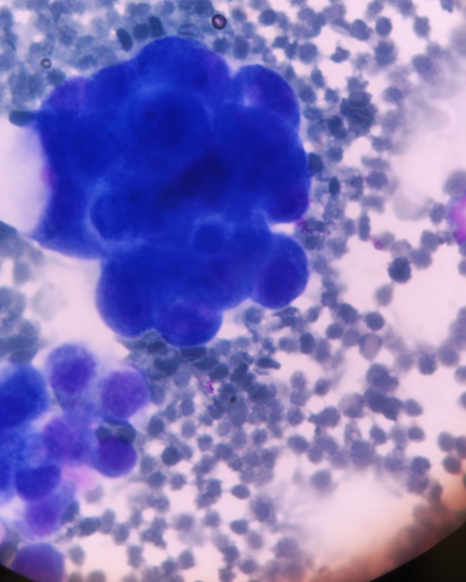
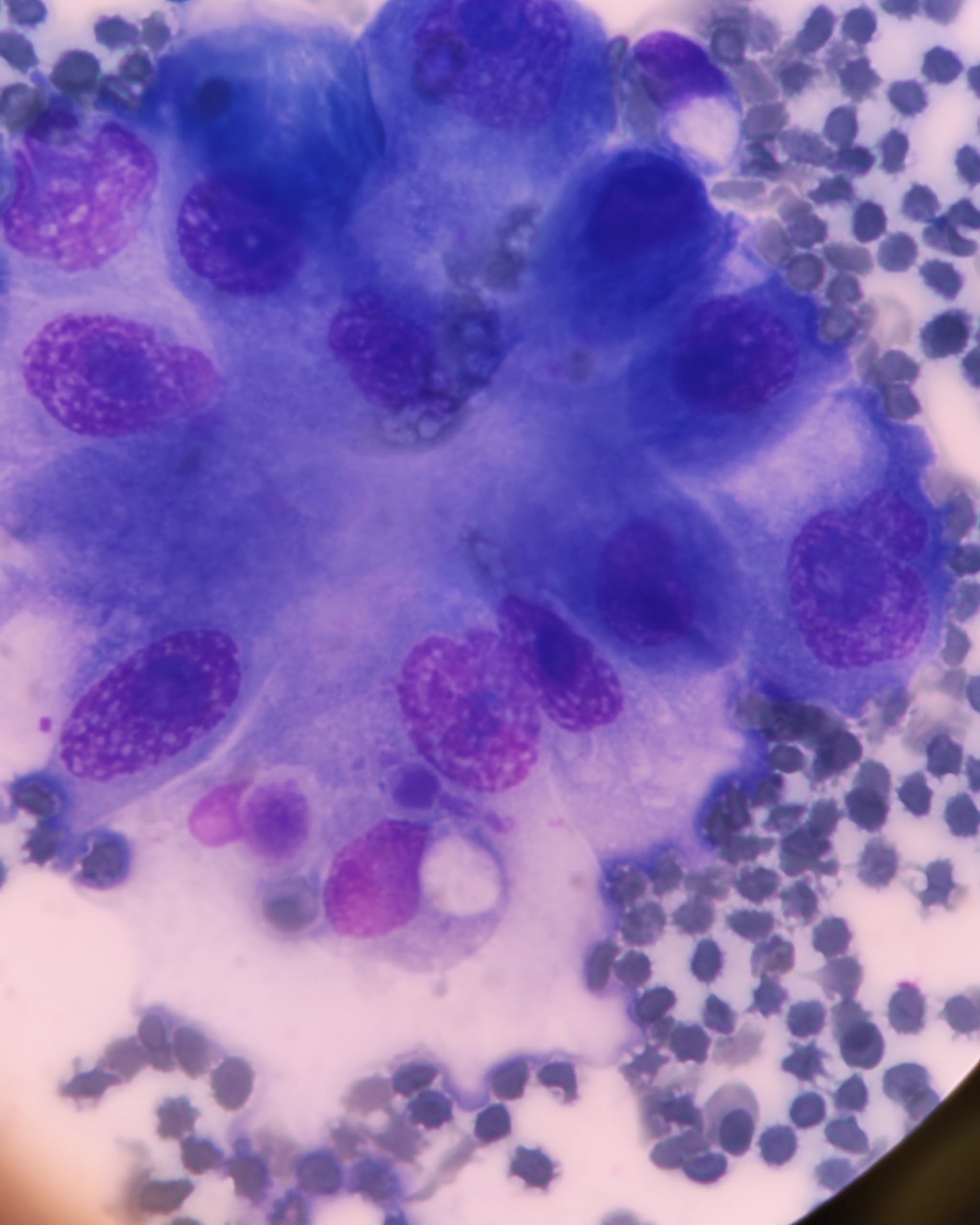
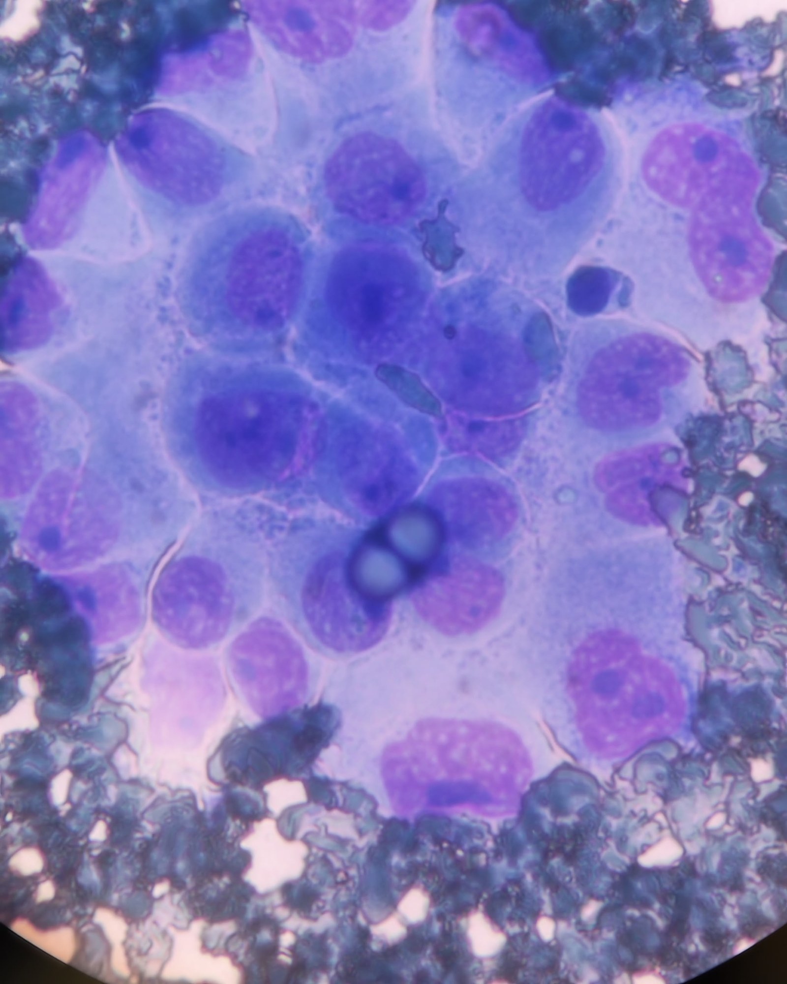
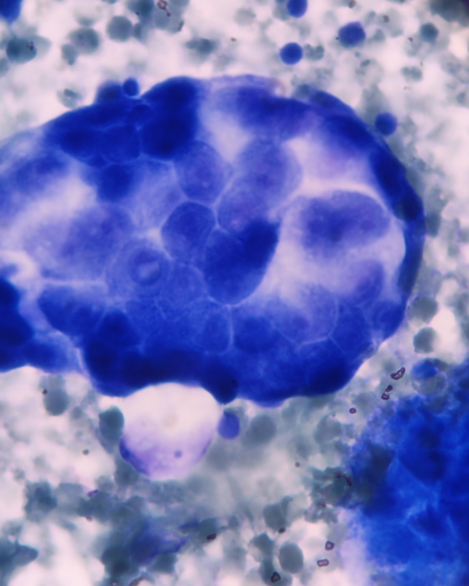
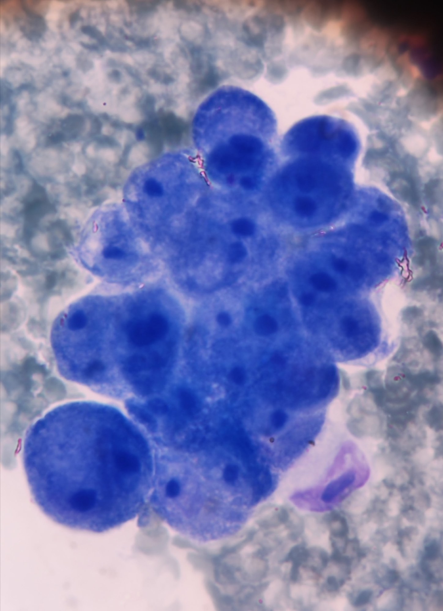
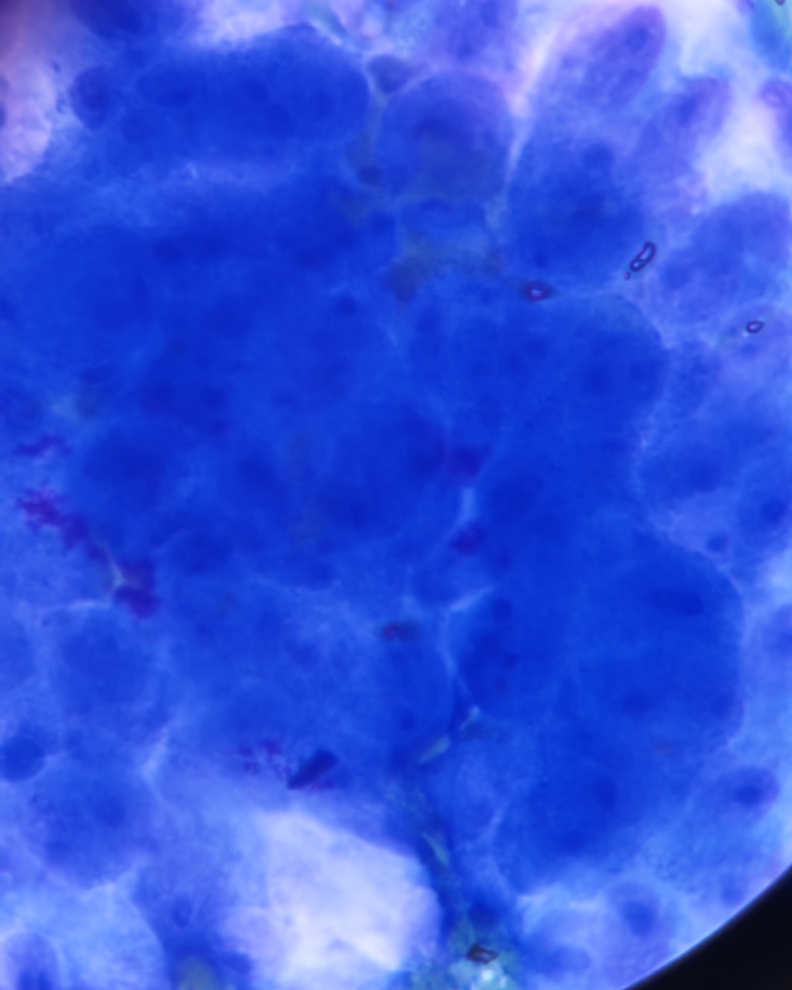
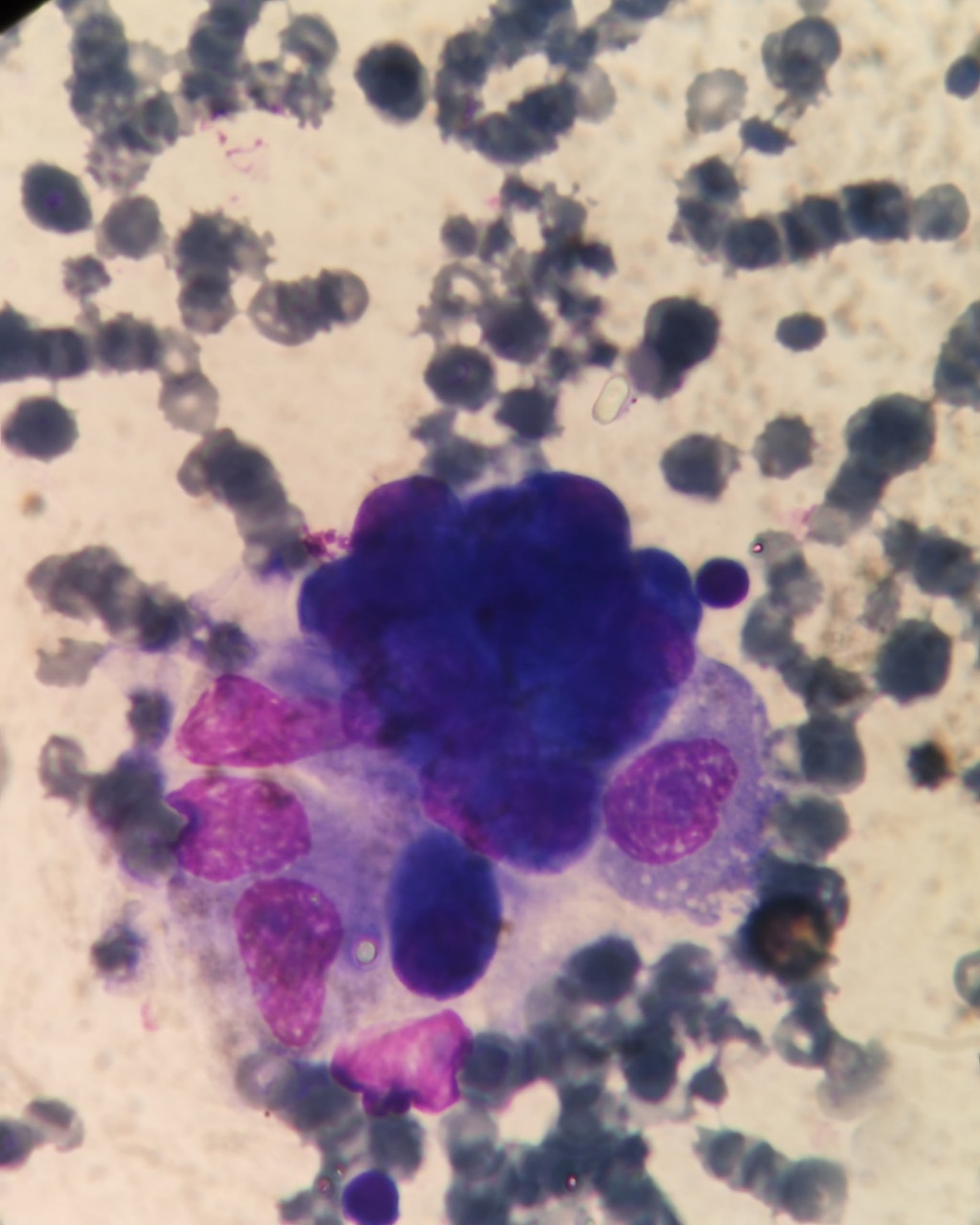
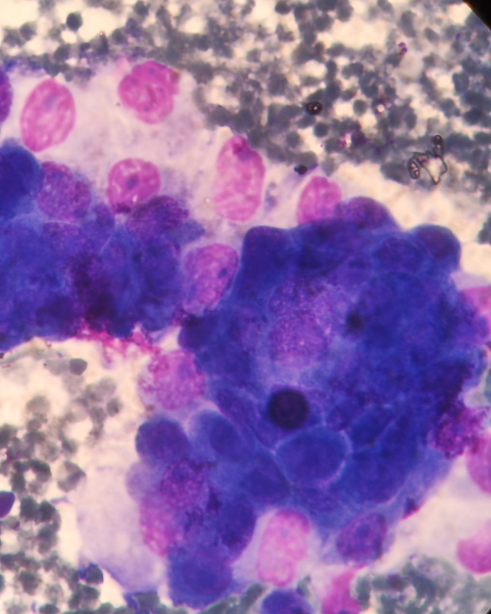
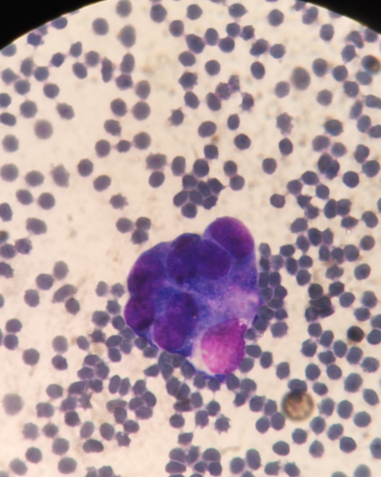
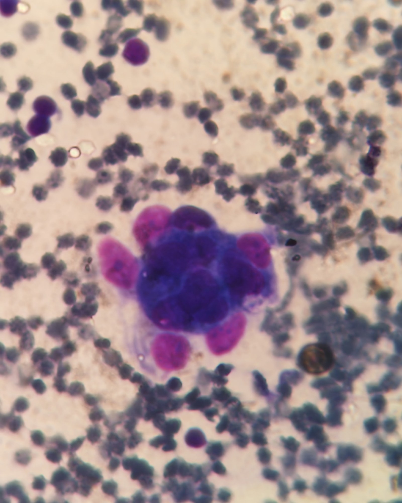
Material - pleural effusion. A patient was admitted to the department when pleural effusion was detected during a routine fluorography. She does not smoke, has never smoked, and underwent fluorography annually with no pathology. Complaints included prolonged dyspnea on physical exertion and lower limb edema. MSCT showed a picture of left-sided pleural effusion; a blastomatous process was not excluded. Pleural puncture was performed, and 700 ml of sero-hemorrhagic exudate was obtained. The preparation showed abundant cellular composition, with cells predominantly arranged in papillary structures and sheets, nuclei displaced to the periphery, and prominent nucleoli.
🙏 The publication was kindly provided by Ekaterina Nozdrina, Omsk.


