

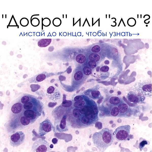
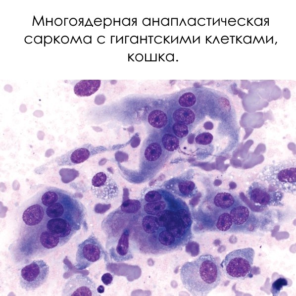
🔬 Cytology. How to distinguish a benign tumor from a malignant one?
Benign tumors usually consist of uniform, well-differentiated cells. However, diversity in cell size, shape, and behavior is an alarming sign. Below are the main signs of malignancy that pathologists and cytologists pay attention to: 🧬 Cellular features:
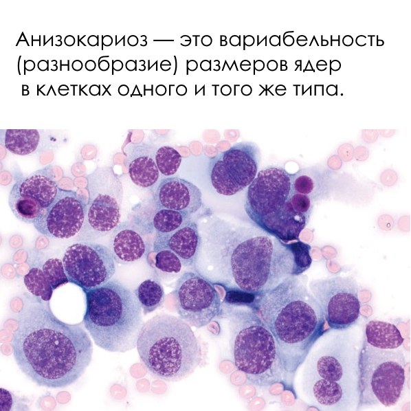
- Different cell sizes (anisocytosis)
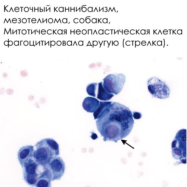
- “Cannibalism” — one cell “eats” another
- Poor differentiation — cells lose the characteristics of their “native” tissue
🧫 Cytoplasm:
- Little cytoplasm compared to the nucleus (high nucleo-cytoplasmic ratio)
- Strong basophilia — cells stain intensely
🧠 Nucleus:
- Different nuclear sizes (anisokaryosis)
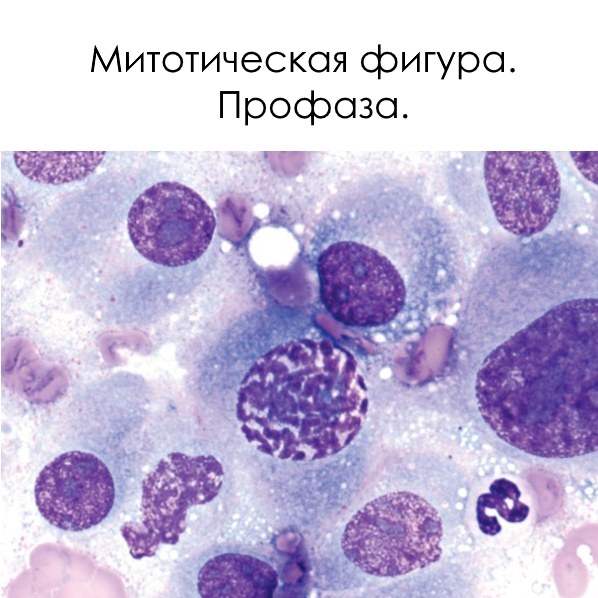
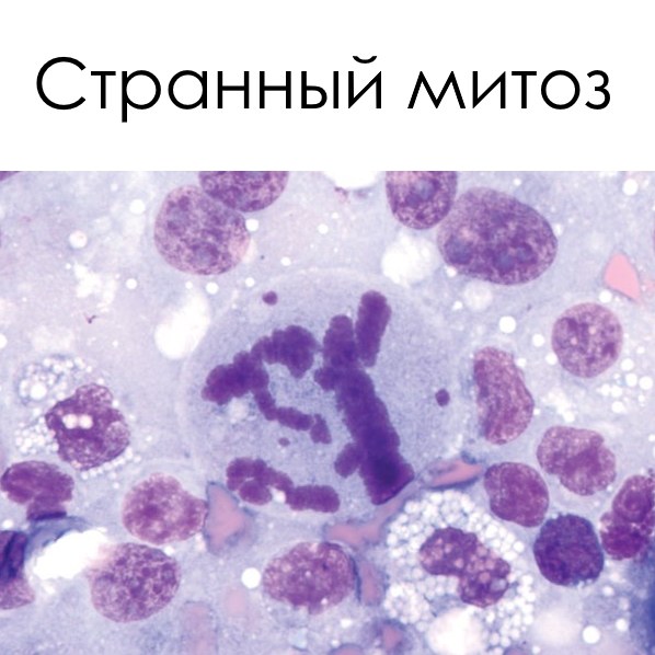
- Atypical mitoses (cell divisions)
- Multinucleation
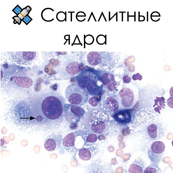
- “Satellite” nuclei
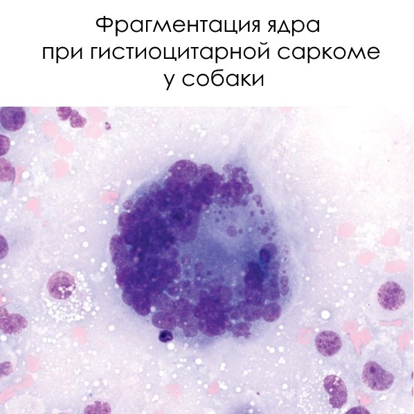
- Fragmentation, hyperchromasia, nuclear crowding
🔎 Nucleoli:
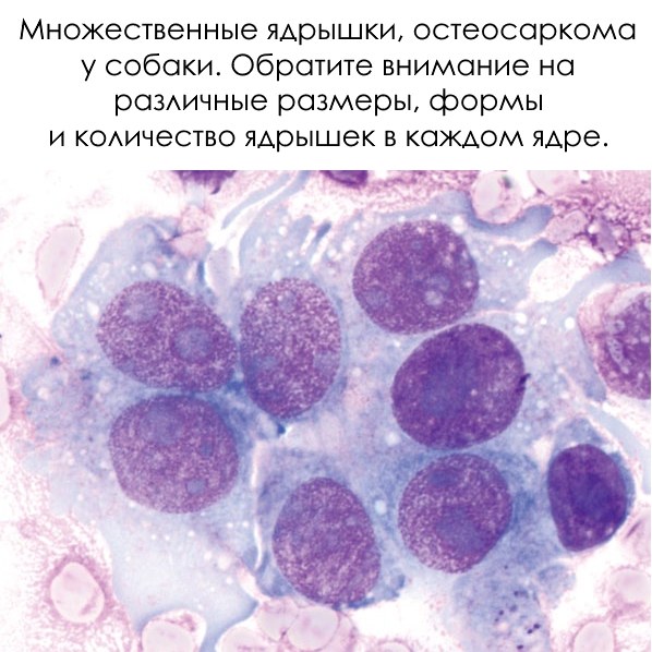
- Large and/or multiple
- Various in shape and size
⚠ It is especially important to carefully examine cell nuclei — most signs of malignancy are associated with them. The more mitoses (especially atypical ones) — the higher the risk that we are dealing with a tumor.
❗ Cytology lesson. Rare findings from Diagnostic Cytopathology journal →.
🔬Image source: Andrew G. Burton - Clinical Atlas of Small Animal Cytology, 2018


