

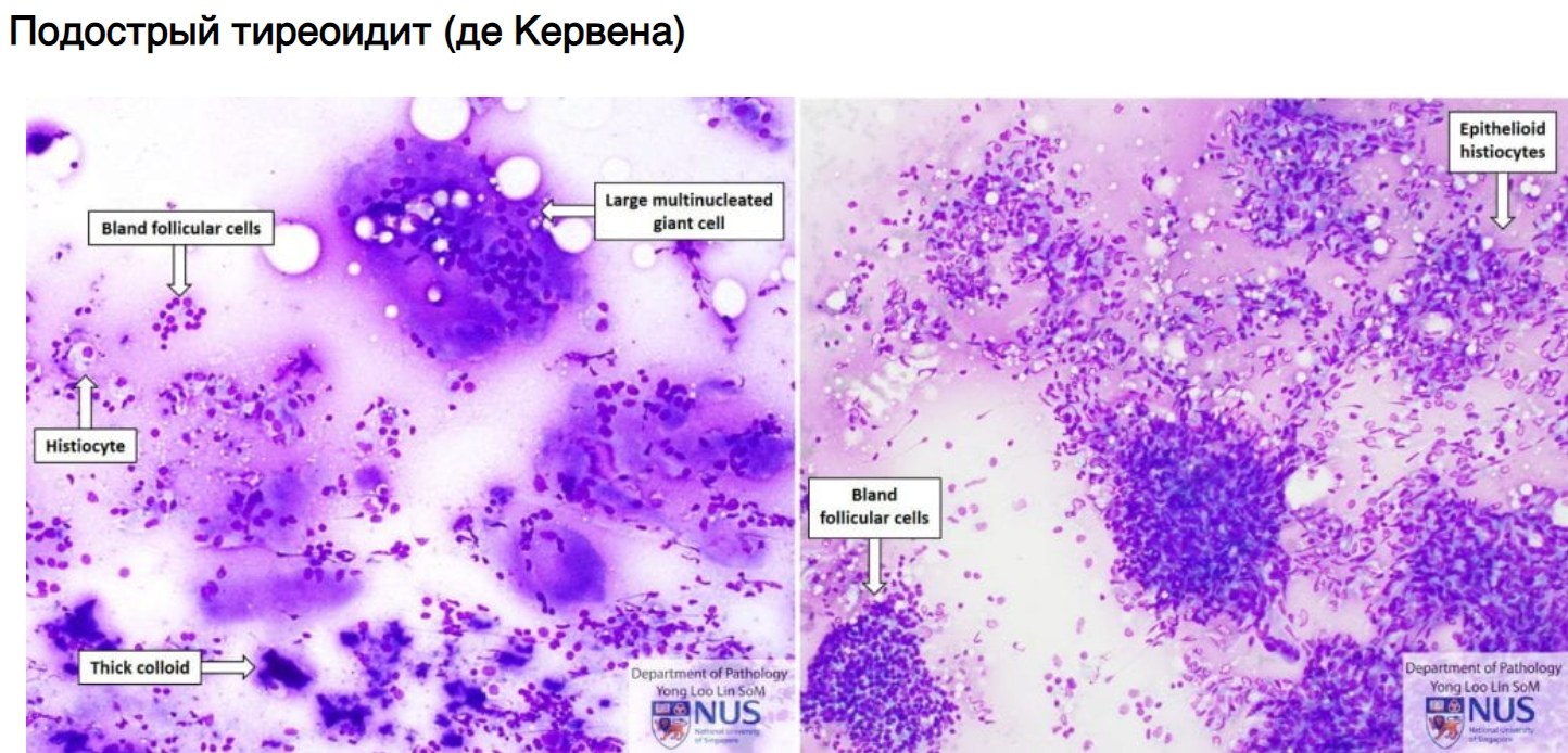
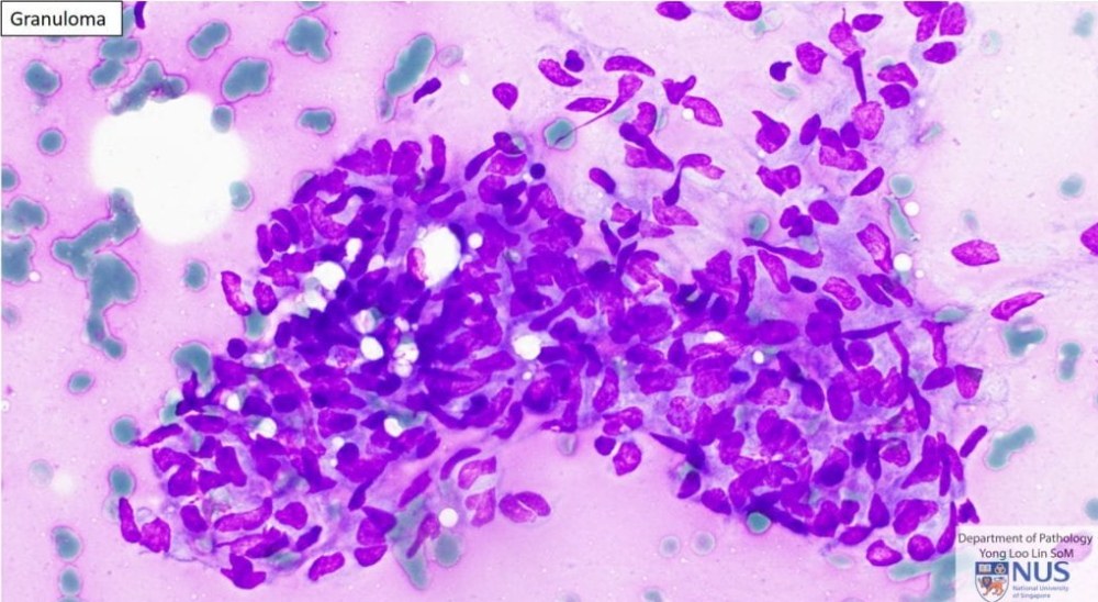
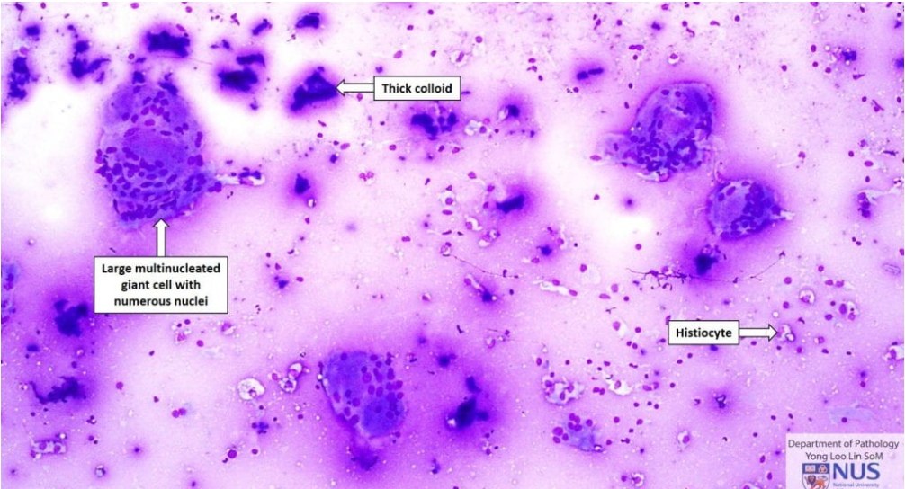
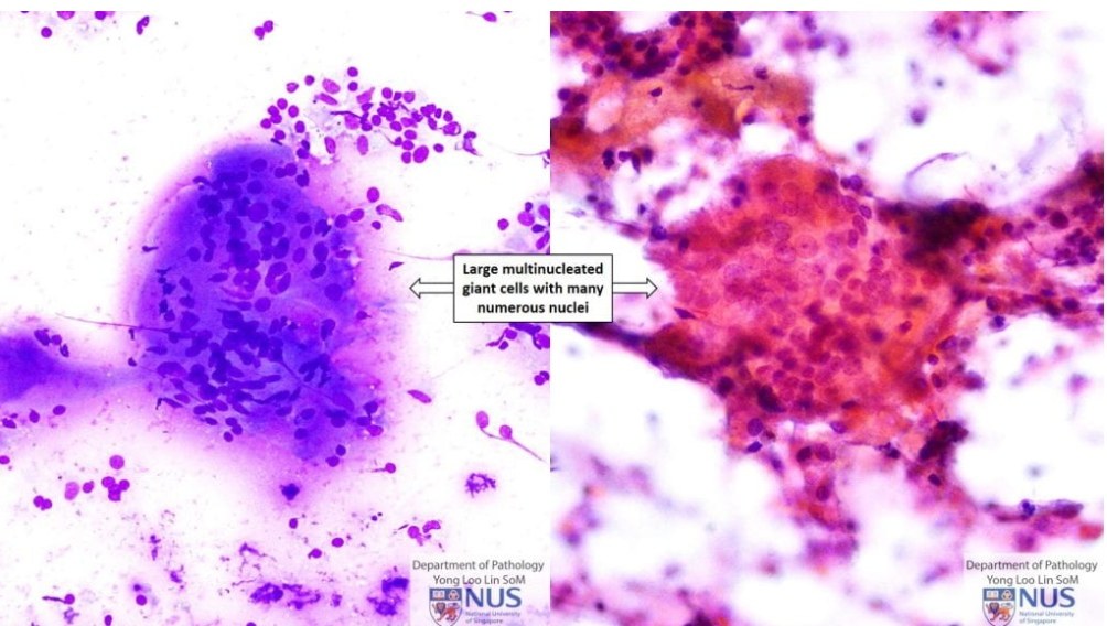
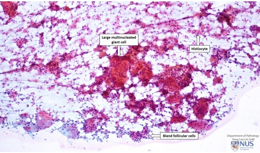
🔬CORRECT ANSWER to the cytology case: Morphological picture of subacute thyroiditis (De Quervain’s thyroiditis, giant cell thyroiditis, granulomatous thyroiditis).
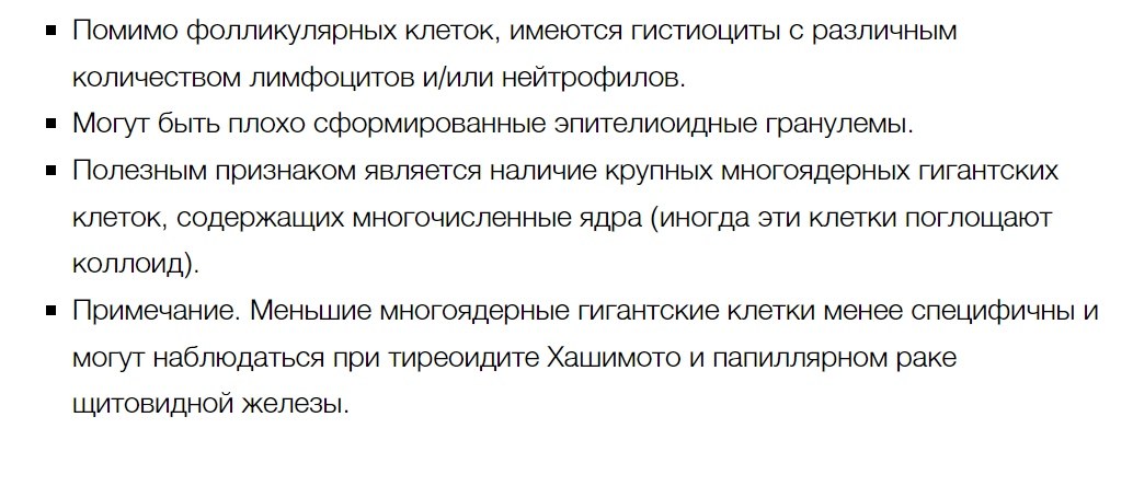
✅In addition to follicular cells, there are histiocytes with varying numbers of lymphocytes and/or neutrophils. Poorly formed epithelioid granulomas may be present. A useful feature is the presence of large multinucleated giant cells containing numerous nuclei (sometimes these cells engulf colloid). ❗Smaller multinucleated giant cells are less specific and can be observed in Hashimoto’s thyroiditis and papillary thyroid carcinoma.
‼See thyroid cytology algorithm →.
👉Source of this case: [cytoweb] - an electronic resource for practical cytopathology.


