

🧪 Ascitic fluid. 👩🏻Woman with a history of ovarian malignancy. Here is such “feathered” atypia in the effusion!
🙏 Photos and description kindly provided by KDL biologist Yulia Guseva, Belorechenskaya Central District Hospital.
Microscopy of ascitic fluid sediment from a patient with ovarian cancer: cytological signs of metastatic involvement and atypical cell complexes.
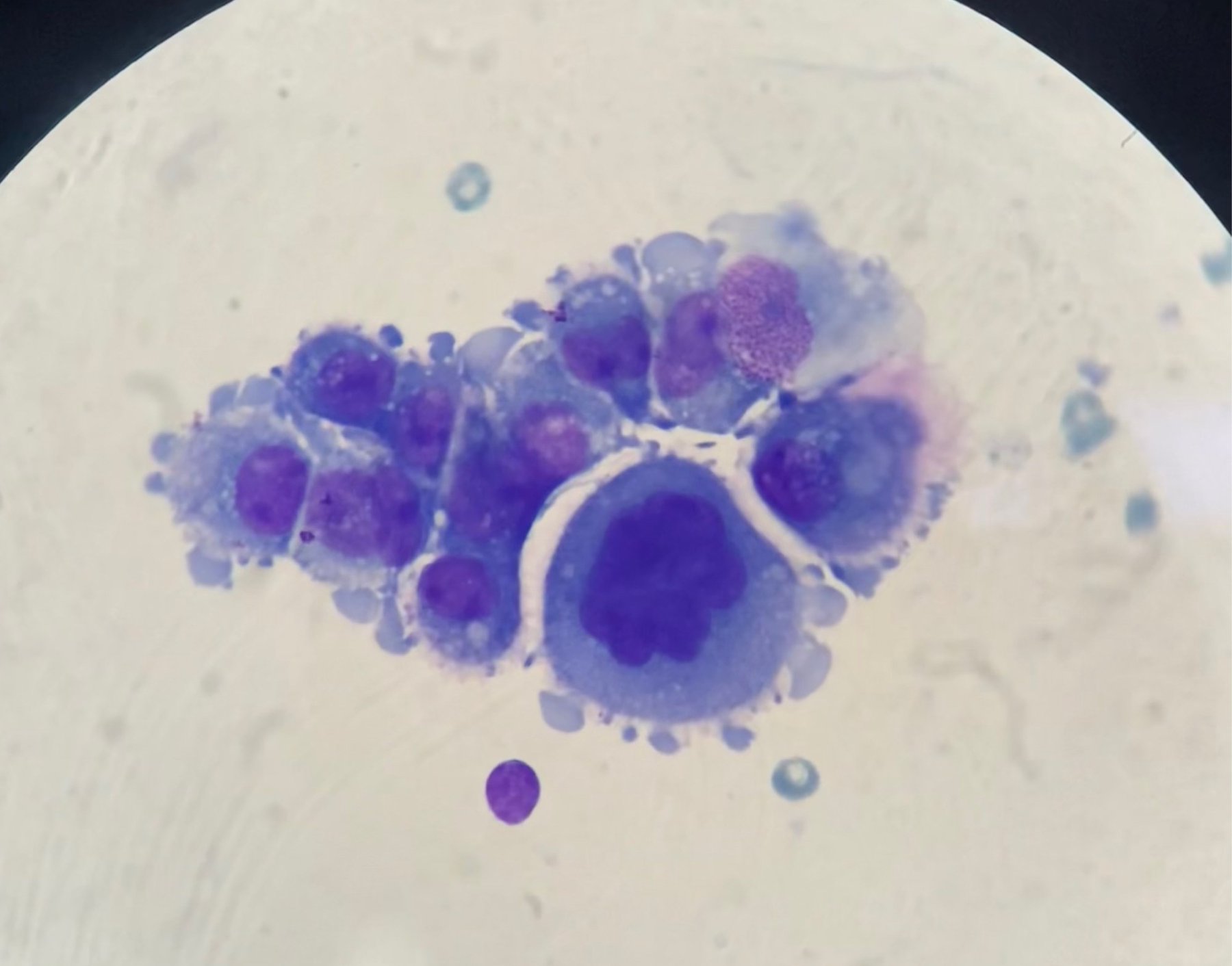
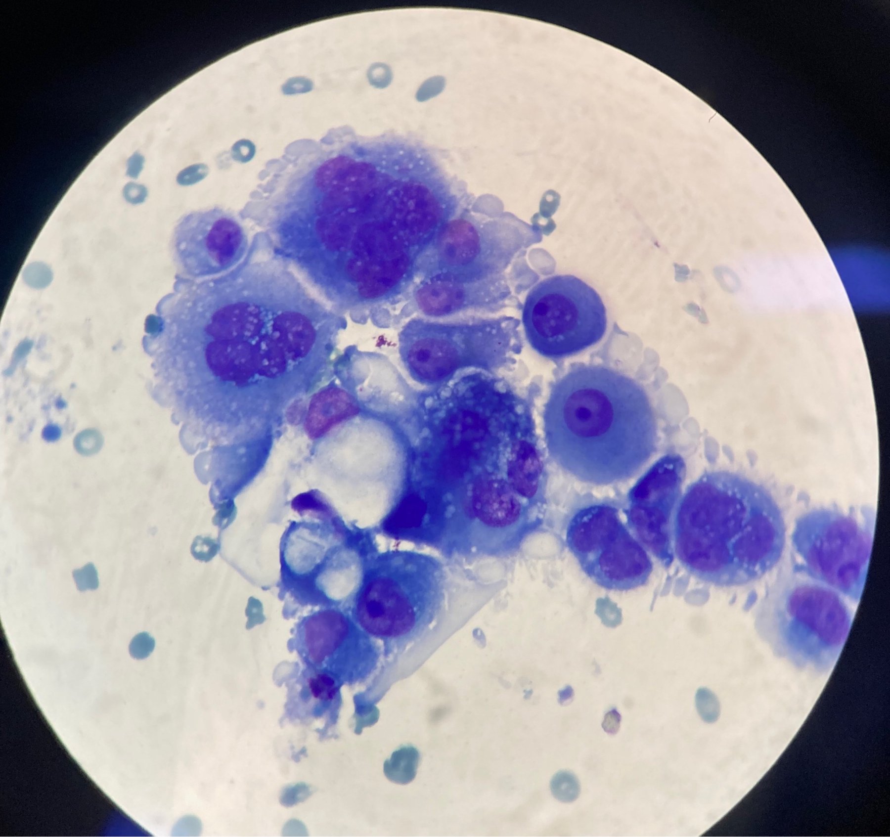
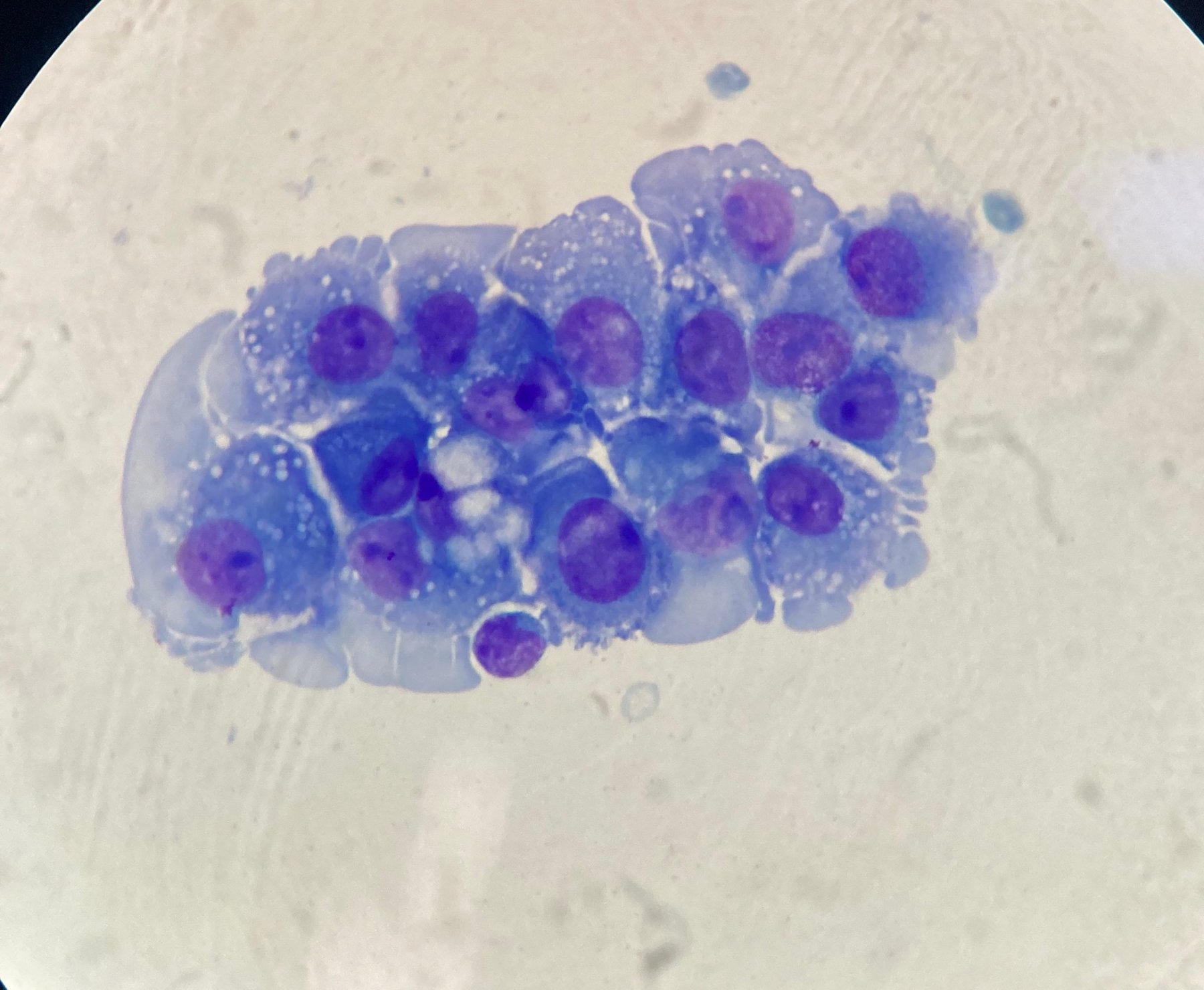
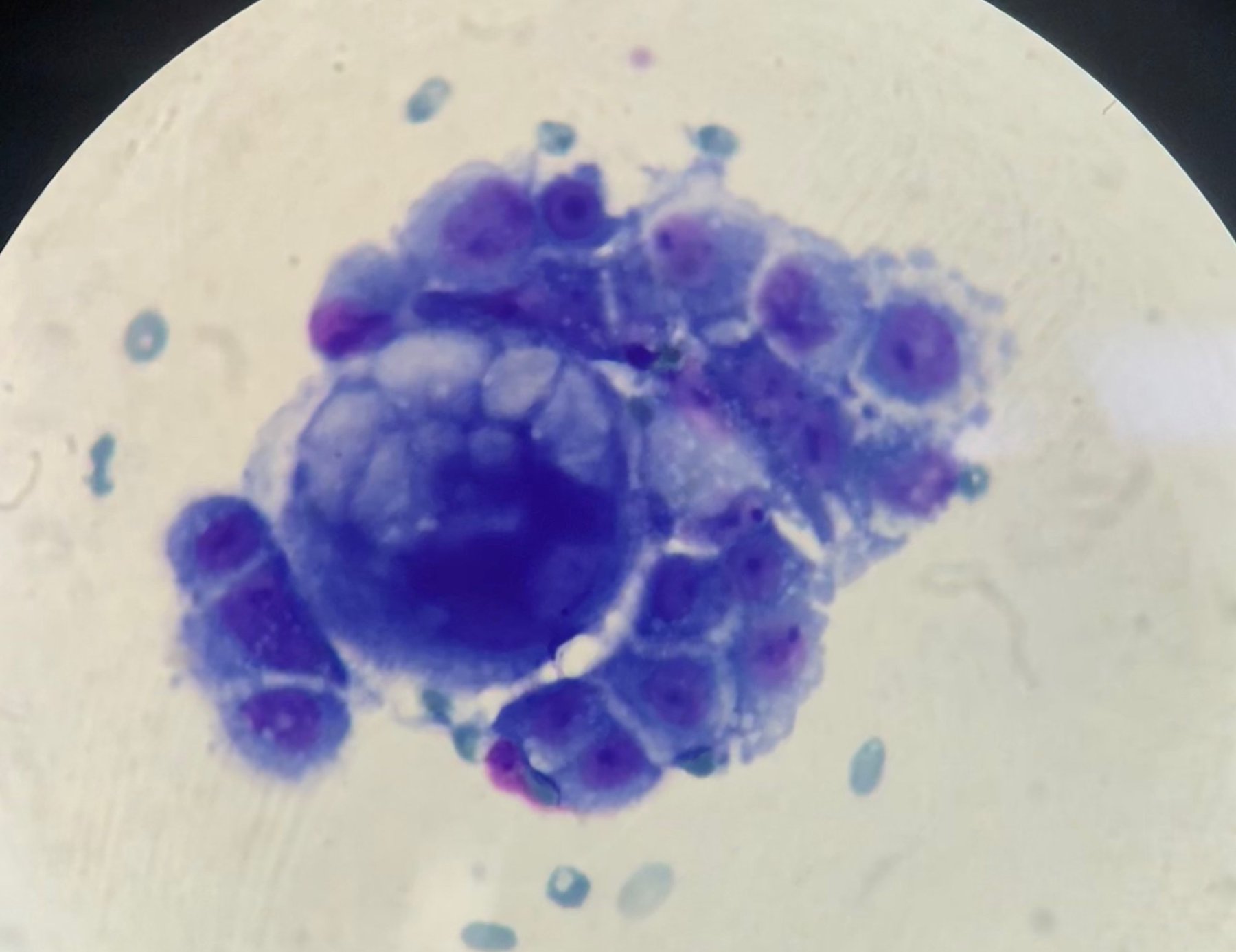
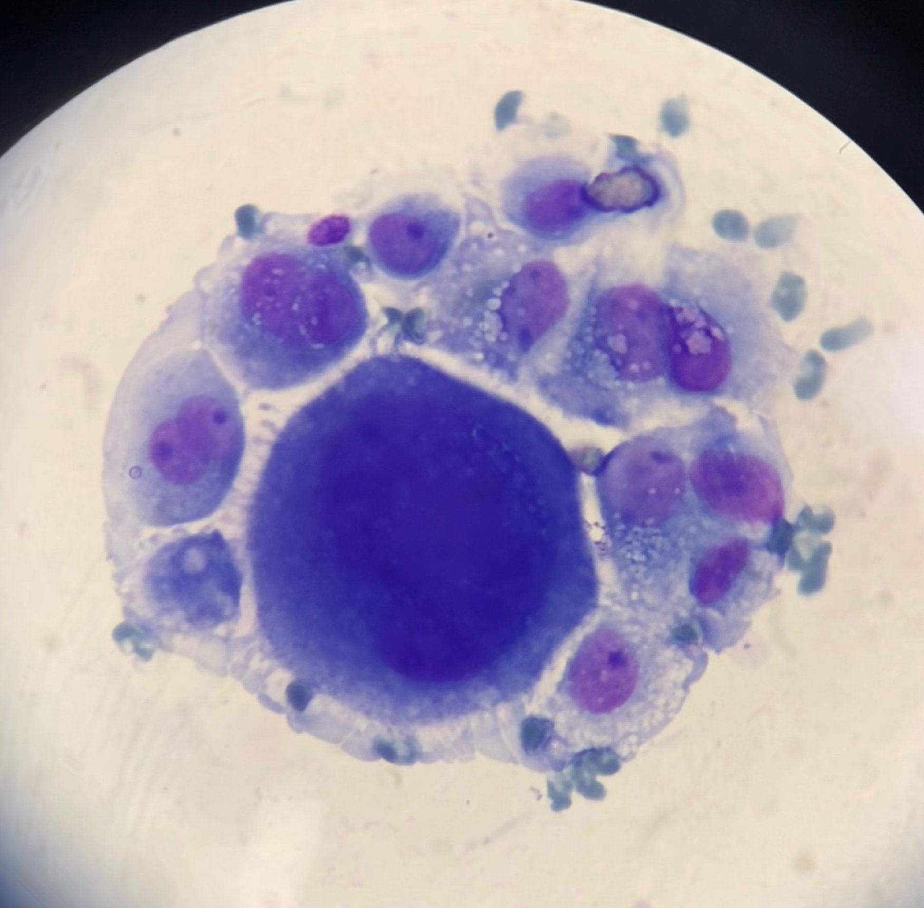
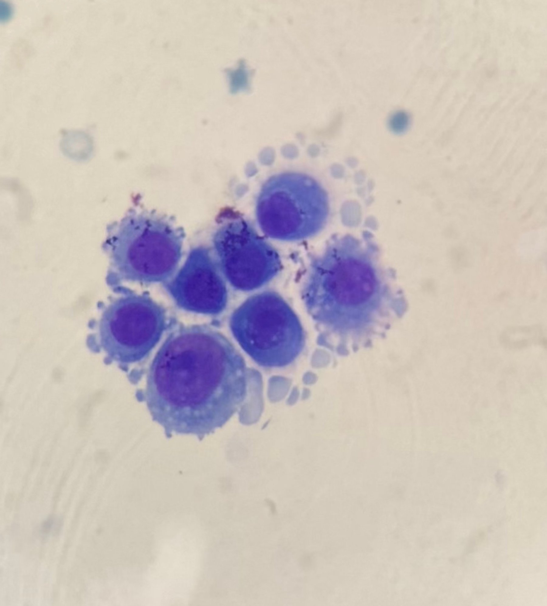
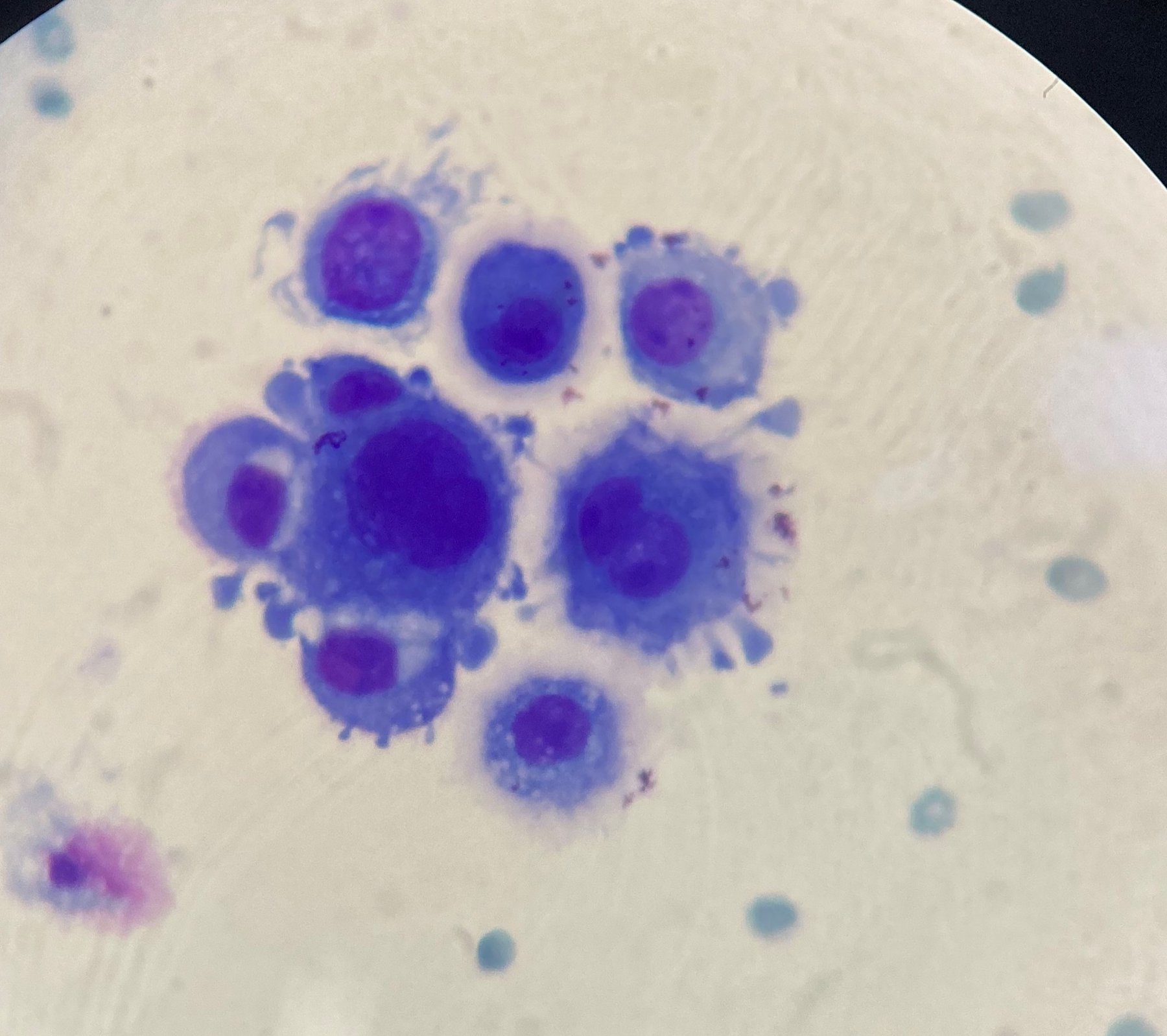
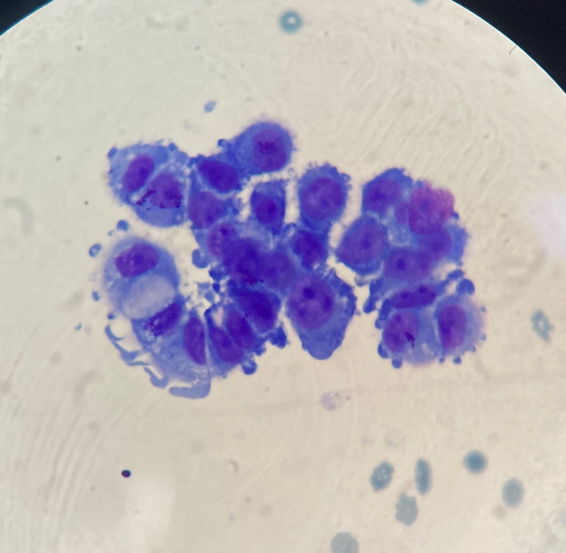
🧪 Ascitic fluid. 👩🏻Woman with a history of ovarian malignancy. Here is such “feathered” atypia in the effusion!
🙏 Photos and description kindly provided by KDL biologist Yulia Guseva, Belorechenskaya Central District Hospital.

A clinical case of bloody sputum cytology: discovery of cell complexes with marked atypia during microscopy with Romanowsky staining.

Cytological findings in ascitic fluid in ovarian cancer. Romanowsky stain: clusters of atypical cells, cellular polymorphism, and vacuolization.

A diagnostic cytology exercise: determine the cause of epithelial changes in a cervical Pap test from a 50-year-old woman. Test your knowledge.