

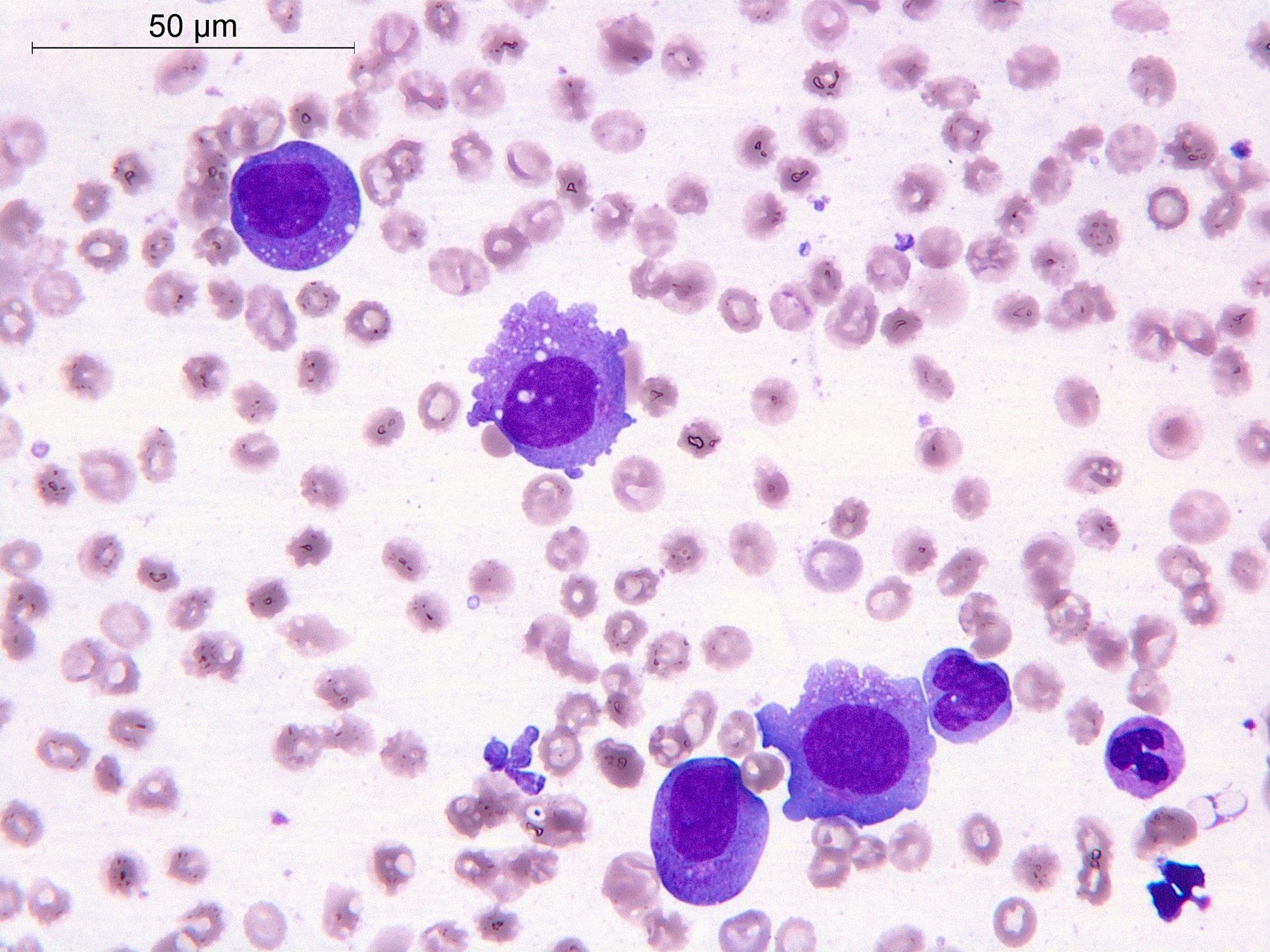
💡 Prize challenge from the Closed Club of Laboratory Hematologists.
❗Male, 66 years old. Diagnosis (based on lymph node biopsy) – nodal lymphoma of cells with follicular T-helper immunophenotype, angioimmunoblastic type. Became ill in January 2023 (B-symptoms, skin rash, superficial lymphadenopathy). Post-6 courses of CHOEP. Complete metabolic response (September 2023). Progression from May 2024. Repeat lymph node biopsy with the same diagnosis. Subsequently received 5 courses according to the GPD regimen. Clinical and hematological remission achieved.
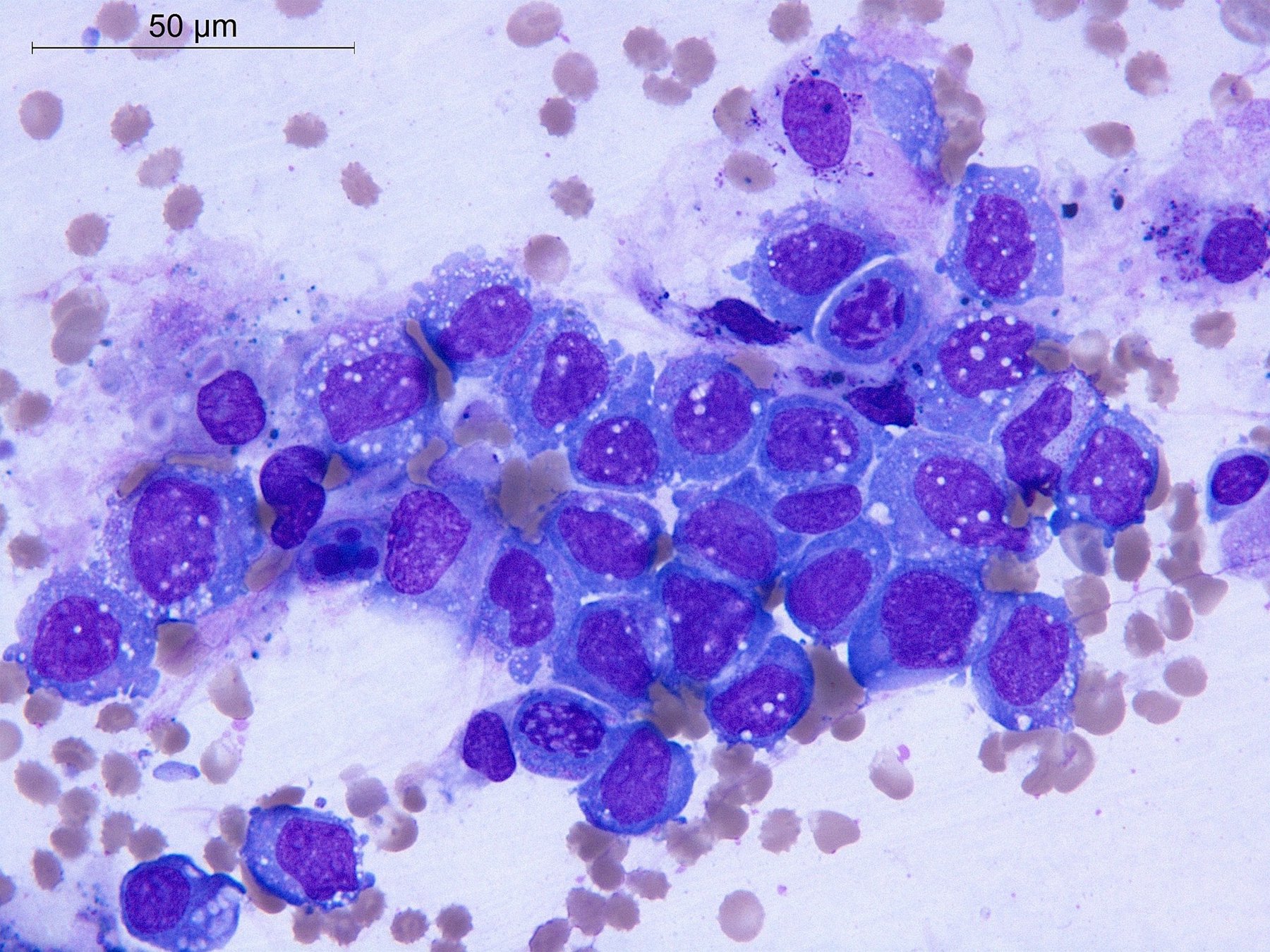
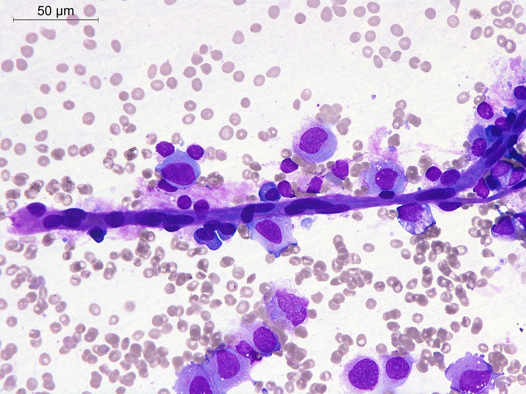
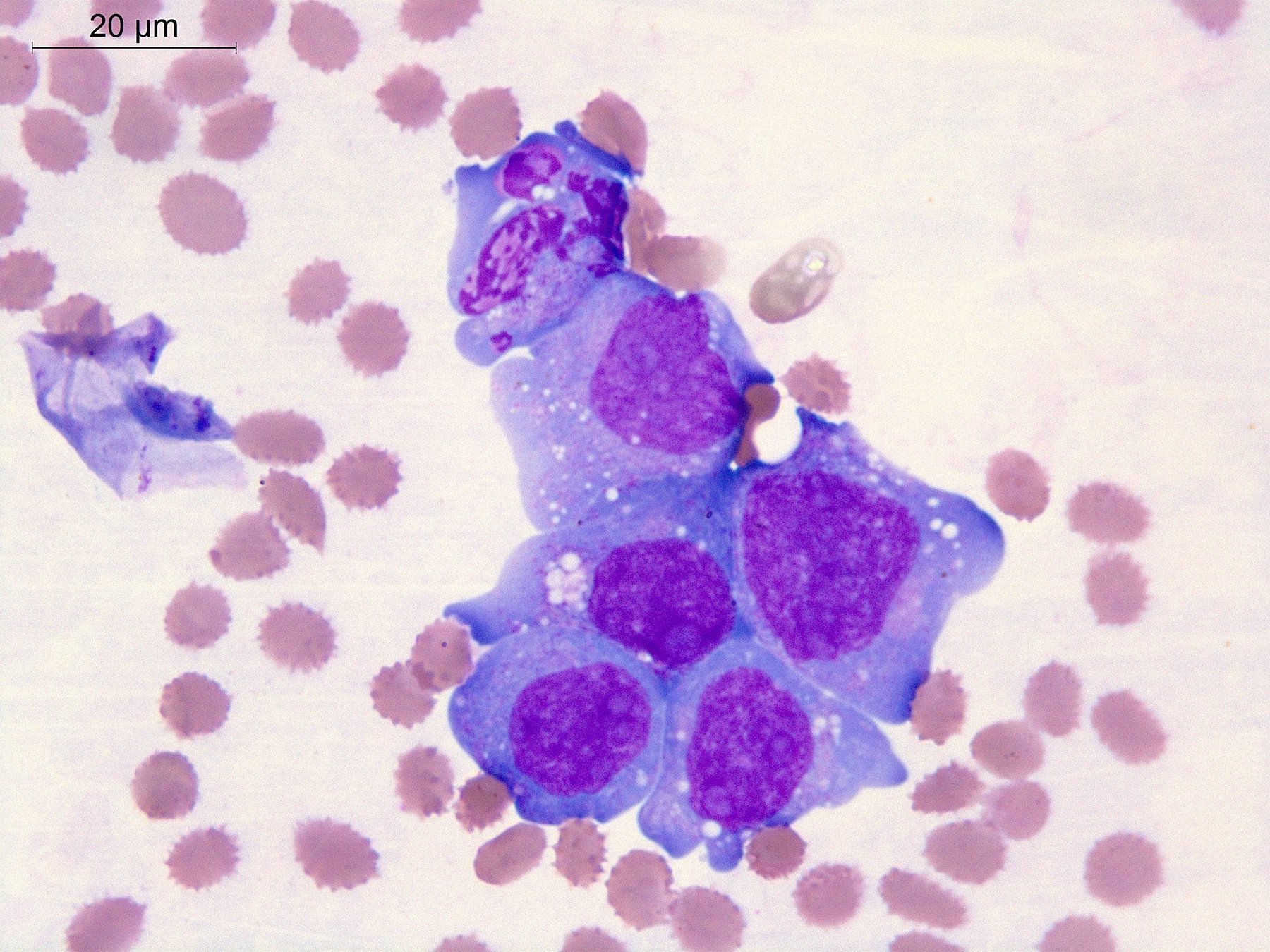
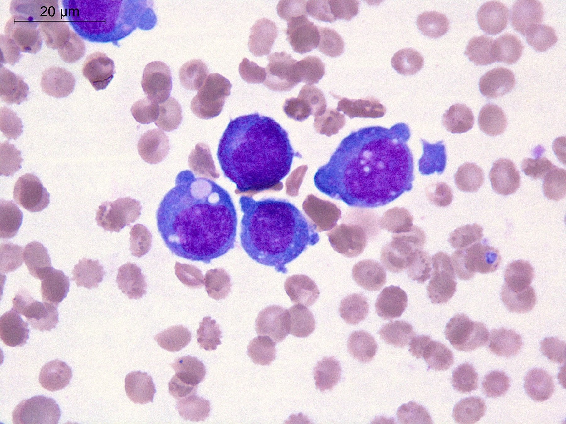
In November 2024, complications arose: 🩸 post-cytostatic neutropenia grade 3, thrombocytopenia grade 4, anemia grade 2. Due to prolonged pancytopenia, a bone marrow aspirate puncture was performed for immunophenotyping (IPT) and a myelogram was conducted.
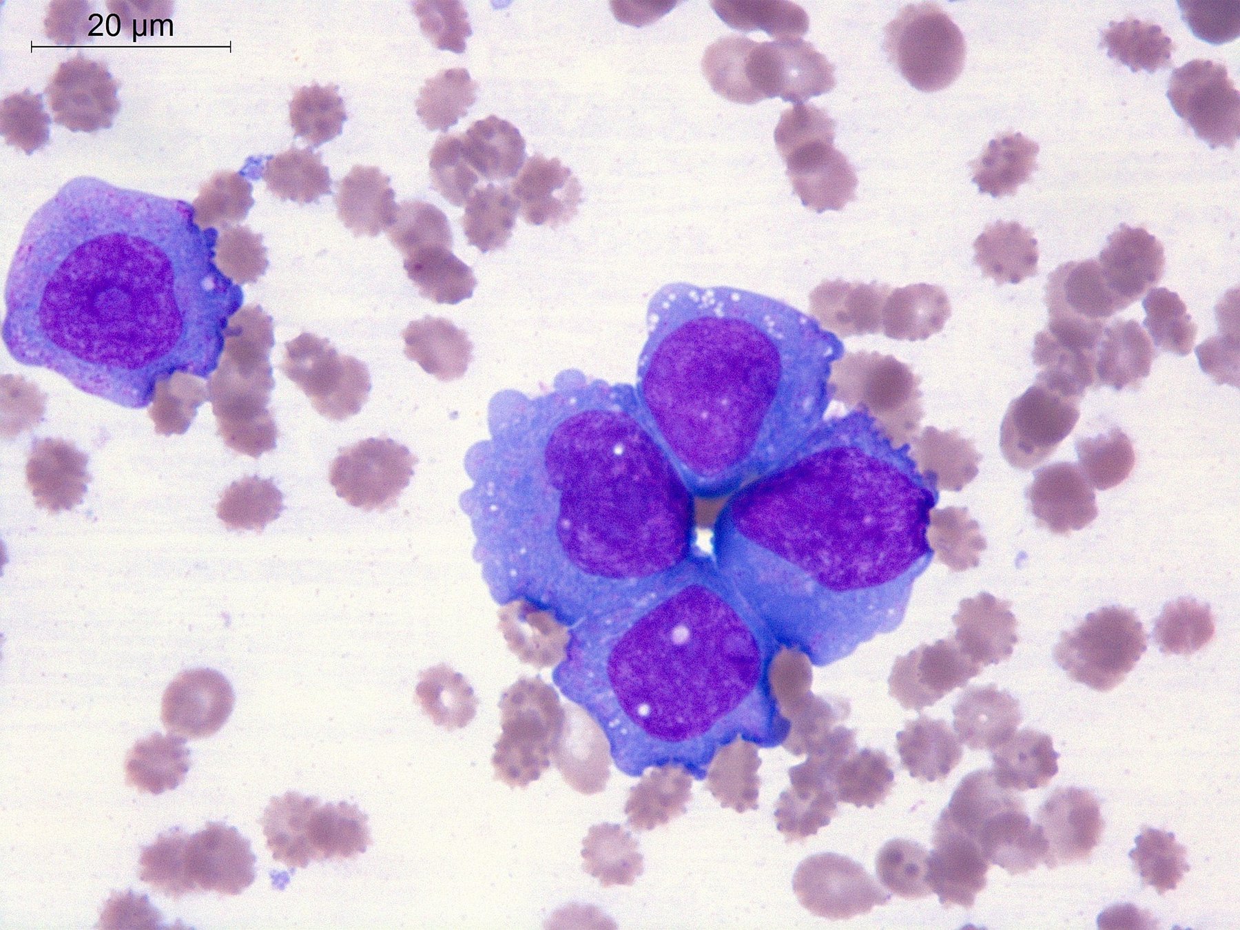
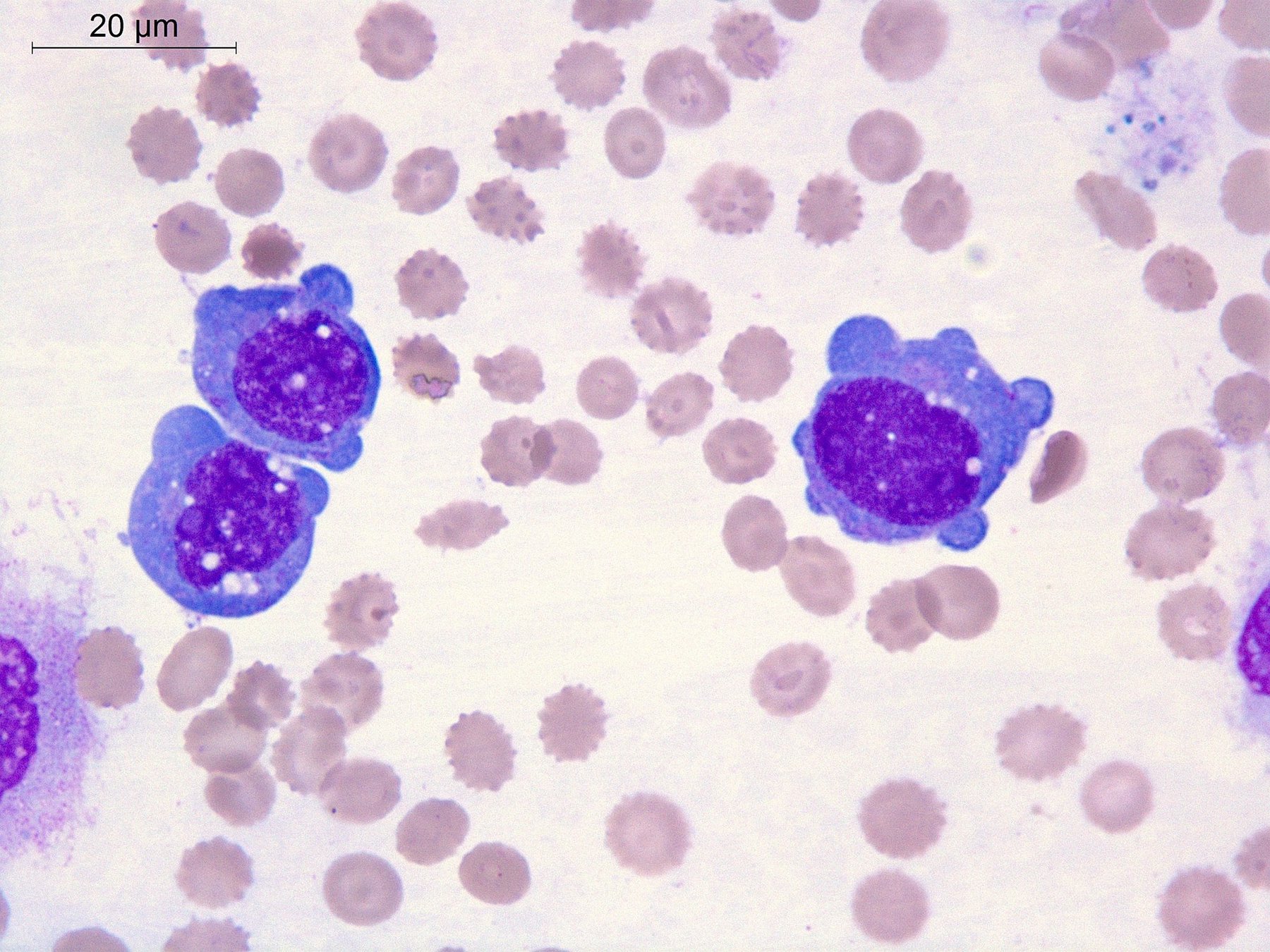
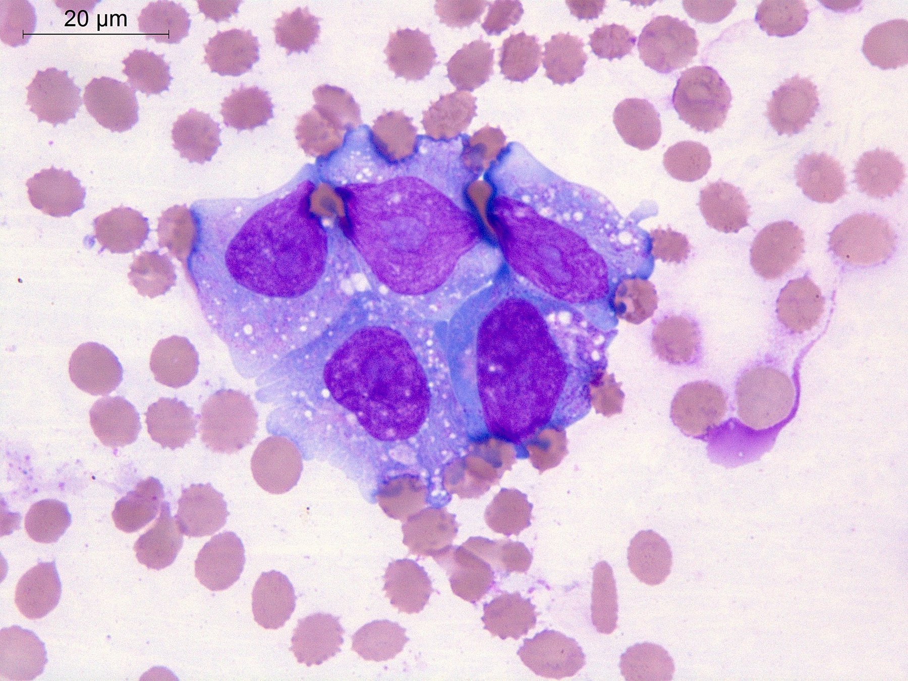
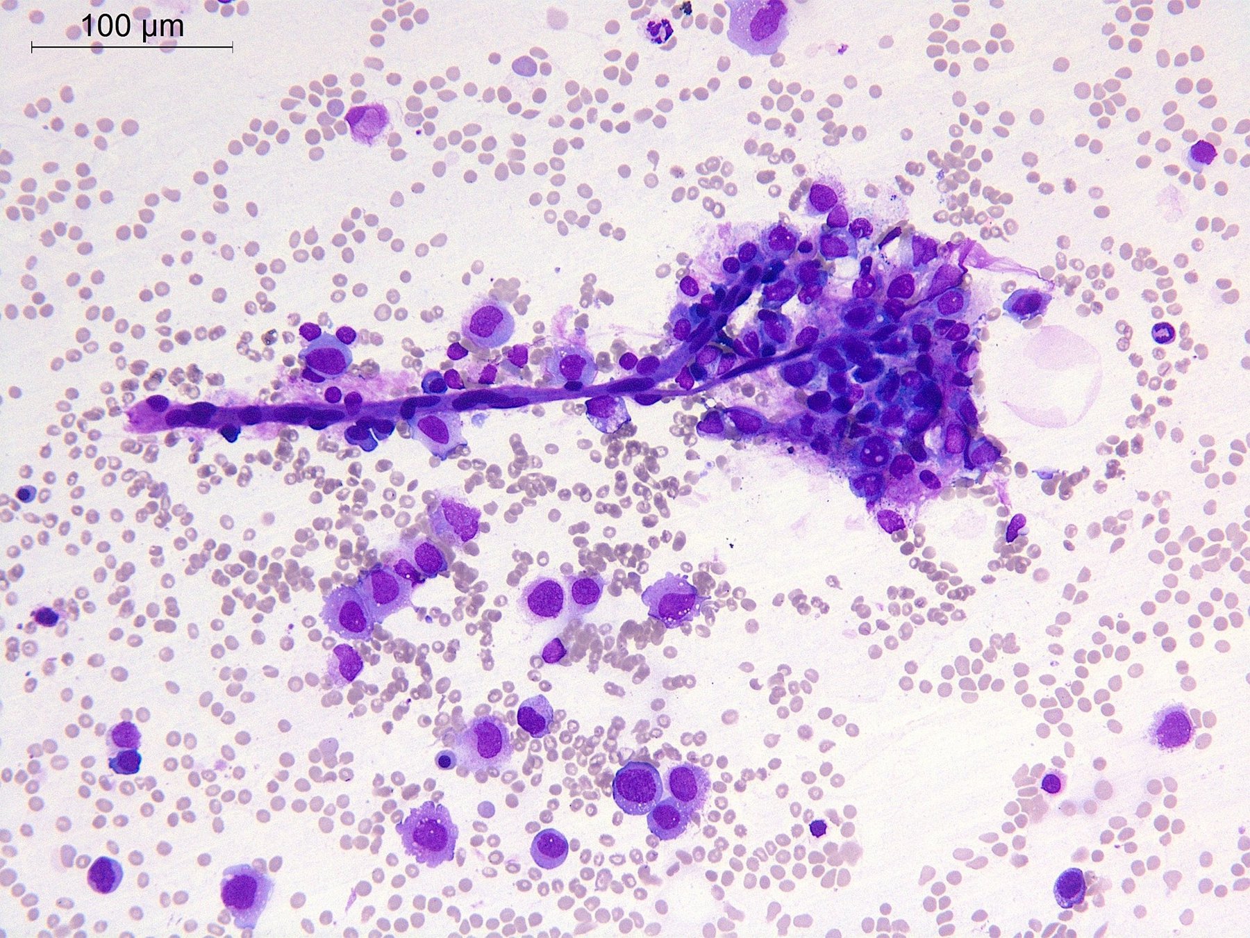
❓We present to your attention cytological preparations of bone marrow aspirate (9 images now + 9 images will be published in the comments later). What diagnosis did you assume based on the cell morphology? Please substantiate your answer, considering the identified cytological features.
🎁 For the first correct answer, a prize: a 30-day Level V subscription to training in our online school! Everyone who also answers correctly in the comments will receive an invitation to the closed club. THE CORRECT ANSWER with IPT results will be published IN 3 DAYS! Participate and win!
👉 The images and task were kindly provided by Elena Belyakova, Clinical Laboratory Diagnostics Doctor, Pathologist, member of the FLM Hematology Committee, Department of Molecular Morphology, E.E. Eichwald Clinic of I.I. Mechnikov Northwestern State Medical University, St. Petersburg.


