

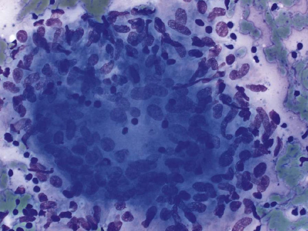
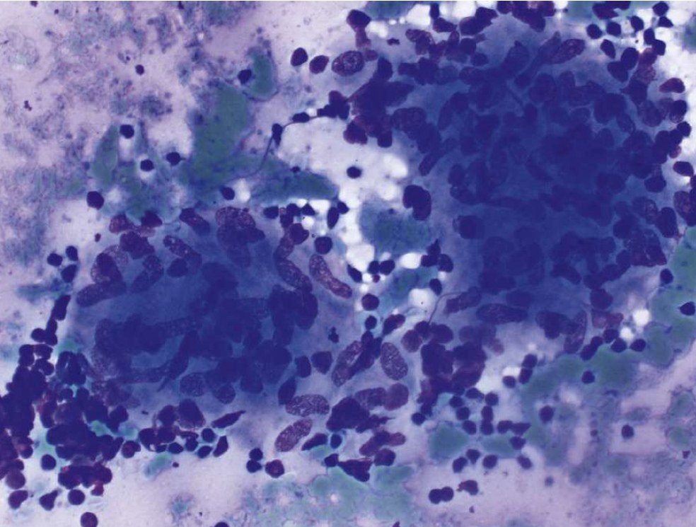
🔬Granulomatous inflammation. Lymph node cytology. ✅Photo №1: This granuloma consists of epithelioid histiocytes and an admixture of lymphocytes. ✅Photo №2: Granulomatous inflammation. There are two non-necrotizing granulomas with epithelioid histiocytes and surrounding small lymphocytes. 👉Diff-Quik stain.
Want to learn more? Visit our LEUKOFORMULA online school website: 🎓LYMPH NODE CYTOLOGY → https://www.leukoformula.ru/2-course-of-study/lymph-node-cytology 🎓LYMPH NODES AND SPLEEN Clinical Cases → https://www.leukoformula.ru/2-course-of-study/lymph-nodes-and-spleen-clinical-cases
Source of post photos: Christopher J. - Atlas of Cytopathology. A Pattern Based Approach, 2020


