

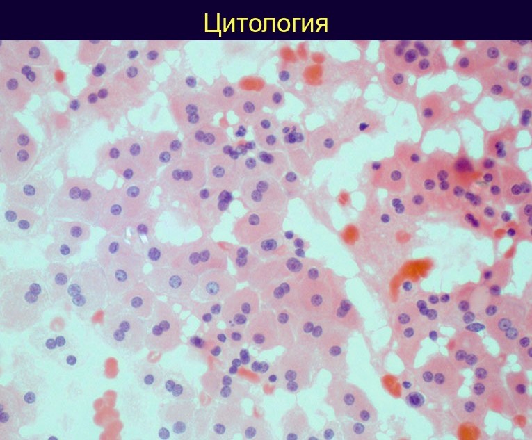
Thyroid cytology. The presented cytological preparation shows a Hurthle cell neoplasm. The cells are characterized by abundant granular cytoplasm and centrally located nuclei, which may contain prominent nucleoli.
Note:
- This morphological picture is consistent with a Hurthle cell neoplasm; however, a definitive conclusion regarding benignity or malignancy requires histological confirmation.
- In 15–45% of cases, a cytological picture suspicious for a Hurthle cell neoplasm turns out to be malignant.
Source: M. Sheaff, N. Singh – Cytopathology: An Introduction (2013). 💎 Find even more useful materials on cytology in our LAB-TESTS library. The link with access to the library is automatically sent to you via private message after paying for a VK Donut Labformula subscription.


