

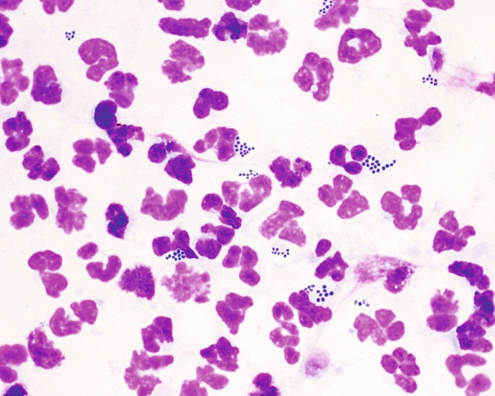
🔬 VETERINARY CYTOLOGY Pyoderma cytology: karyolytic neutrophils with numerous intracytoplasmic cocci.
Localization: Skin (neoplastic skin diseases).
Description: The image shows neutrophils with signs of nuclear degeneration — karyolysis. The nuclei appear swollen and hypochromatic, with a loss of the typical multi-lobed structure. A large number of bacteria (cocci) are observed in the cytoplasm of the neutrophils.
Source: Francesco Albanese - CANINE FELINE SKIN CYTOLOGY SECOND, 2025 Photo source: 🏛 Library “Veterinary Laboratory Medicine”


