

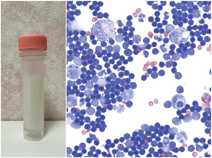
🔬 VETERINARY CYTOLOGY Cat. Pleural effusion. Chyle. (Left) Macroscopically milky appearance. (Right) Predominance of small lymphocytes with a small number of macrophages containing clear punctate vacuoles. Wright-Giemsa stain.
✅ Chylothorax — accumulation of lymph (chyle) in the pleural cavity ☑️ Essence Chyle drains via the thoracic duct into the venous system Contains lymph and chylomicrons (fats from the GI tract) Impaired drainage → accumulation in the pleura
☑️ Causes 🧠 Mediastinal and lung tumors (thymoma, lymphoma) 🔥 Inflammation, fungal granulomas 🫁 Lung lobe torsion ❤️ Heart and pericardial diseases 🩸 Cranial vena cava thrombosis 🧬 Congenital anomalies of the thoracic duct
❓ Idiopathic
💥 Thoracic duct rupture Photo source: 🏛 Library “Veterinary Laboratory Medicine”


