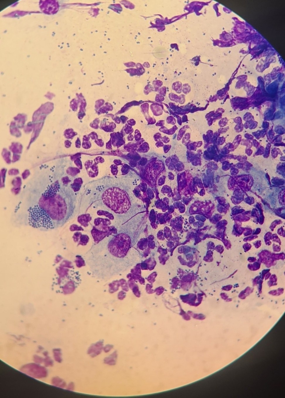
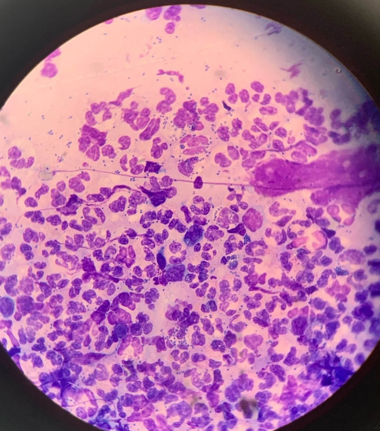
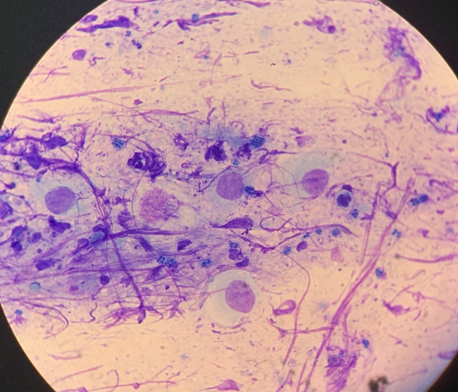
🔬Microscopy challenge for flora smears. ✅The 3 photos show microscopic images of urogenital preparations from 3 different patients, but with the same infection. ❓Which pathogen was detected by PCR analysis? 👉The material for the challenge was kindly provided by clinical laboratory diagnostics physician Oksana Vladislavovna Kadantseva.
Diagnosing Gonorrhea by Microscopic Smear Examination
Microscopic signs of gonorrhea (Neisseria gonorrhoeae) in urogenital smears: clusters of diplococci, leukocyte reaction, and PCR confirmation.