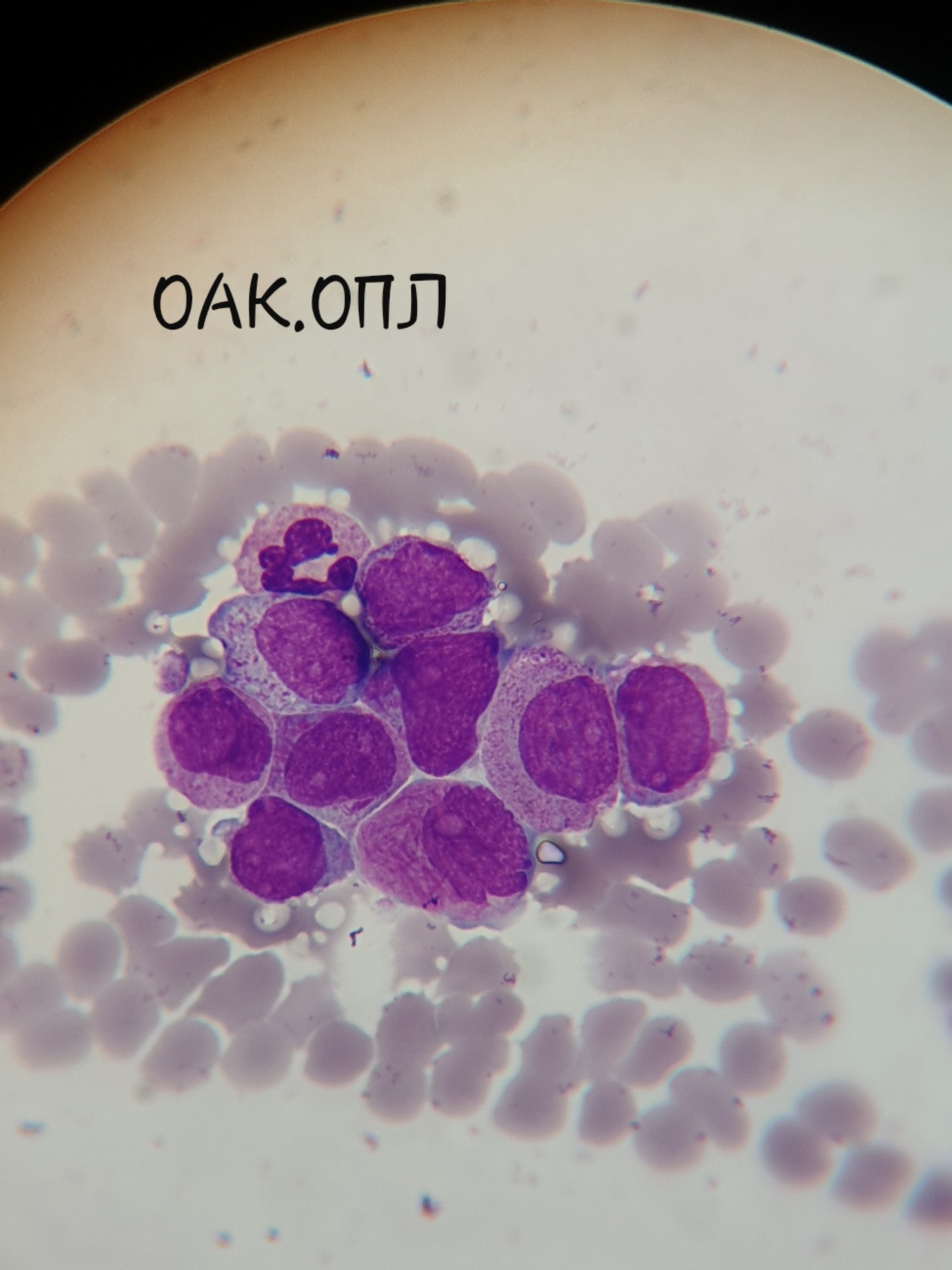
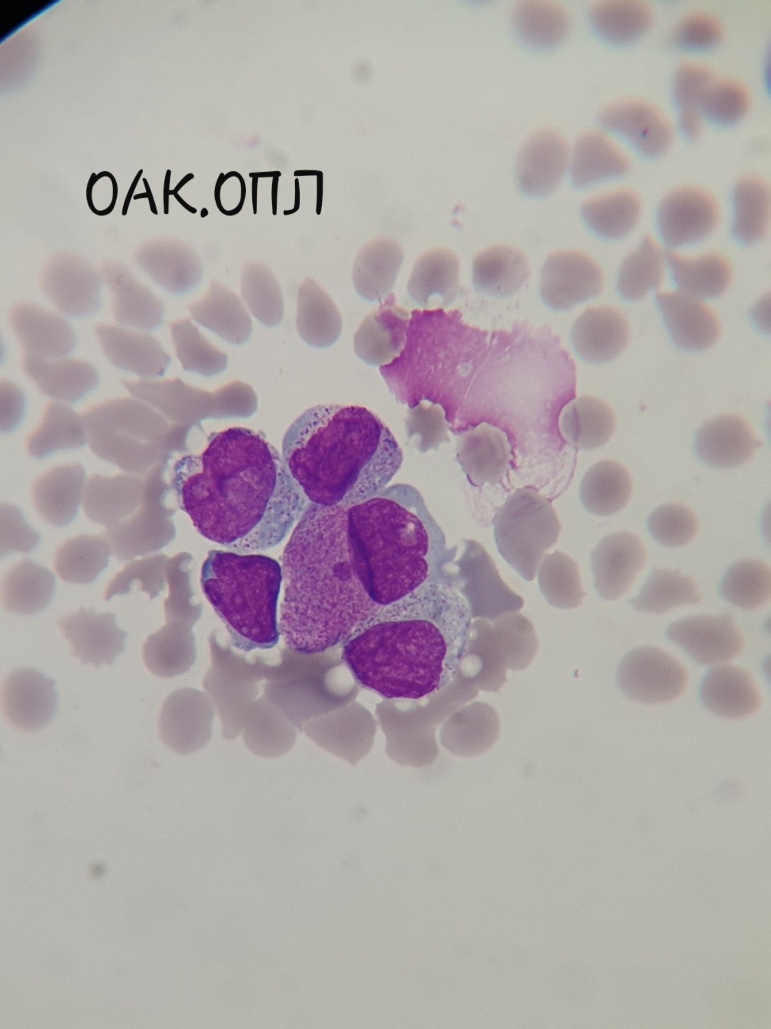
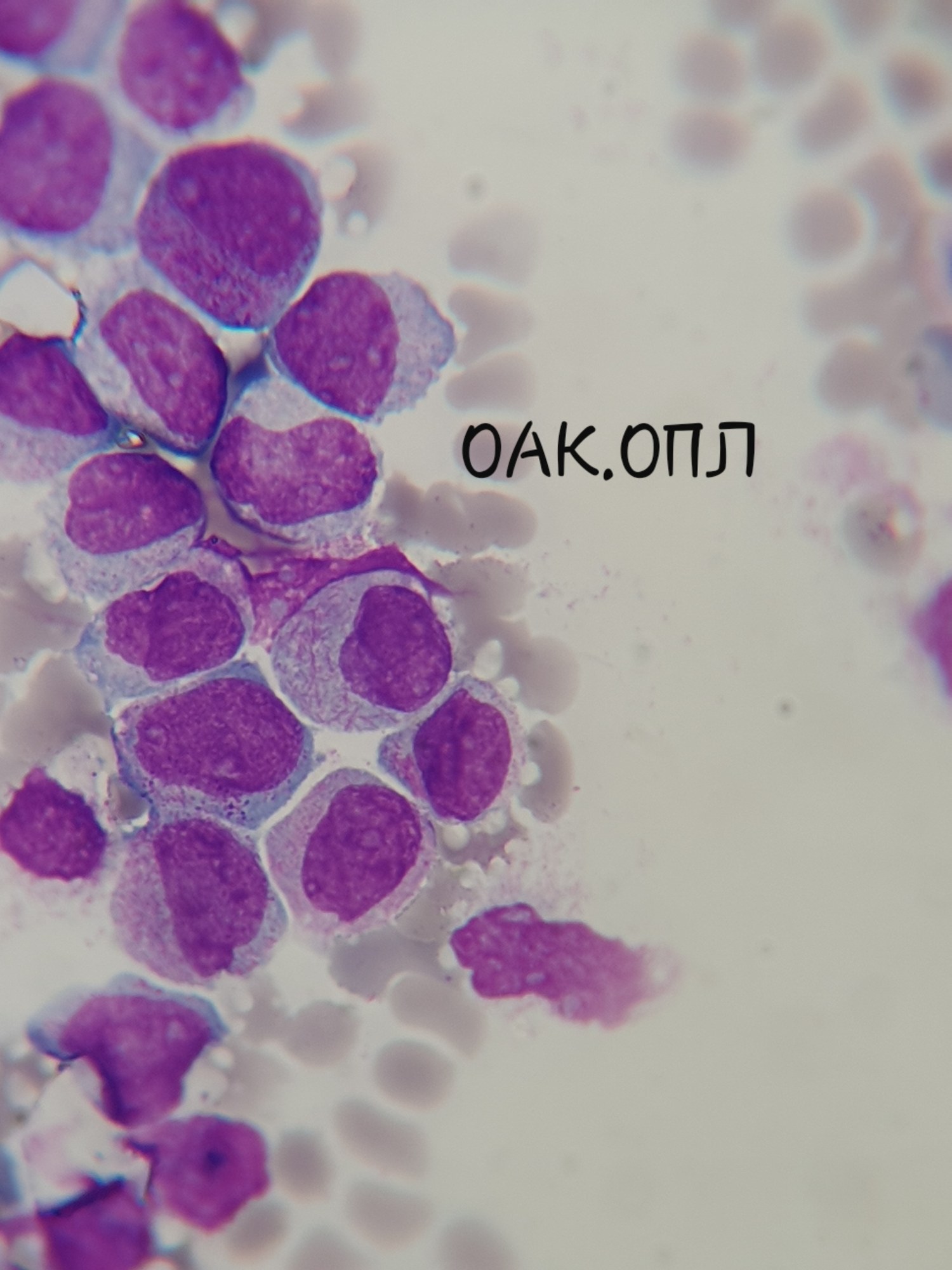
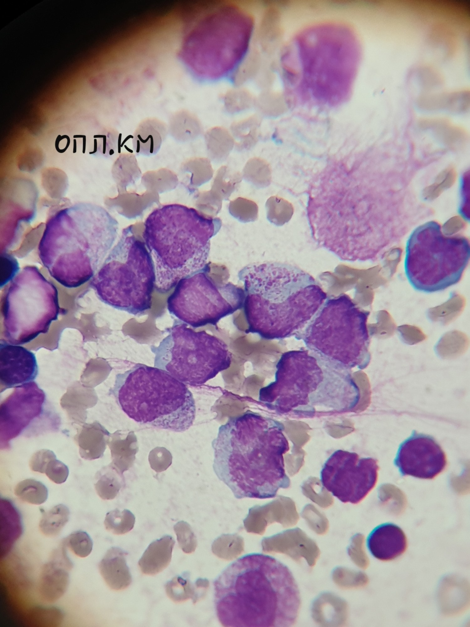
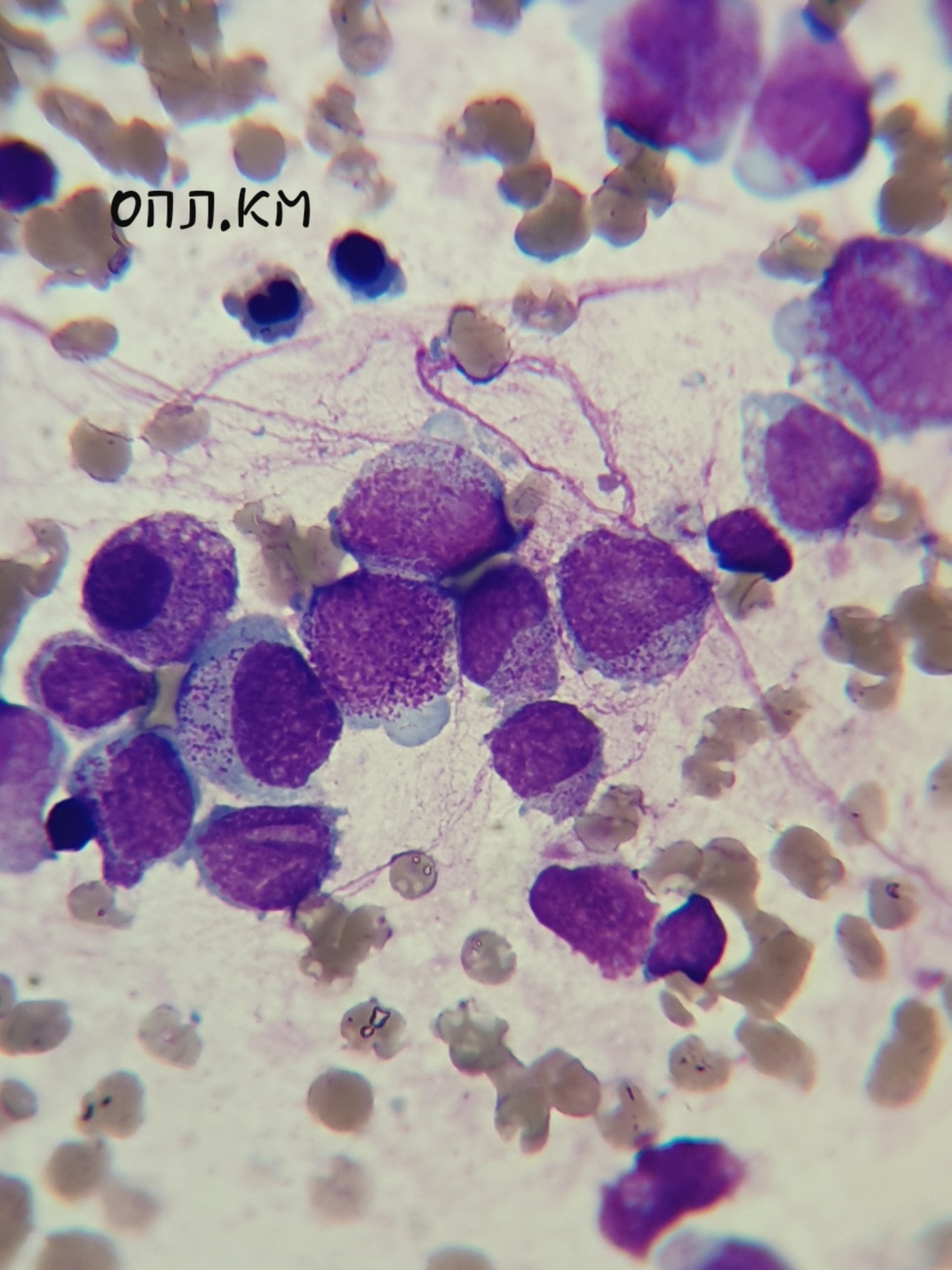
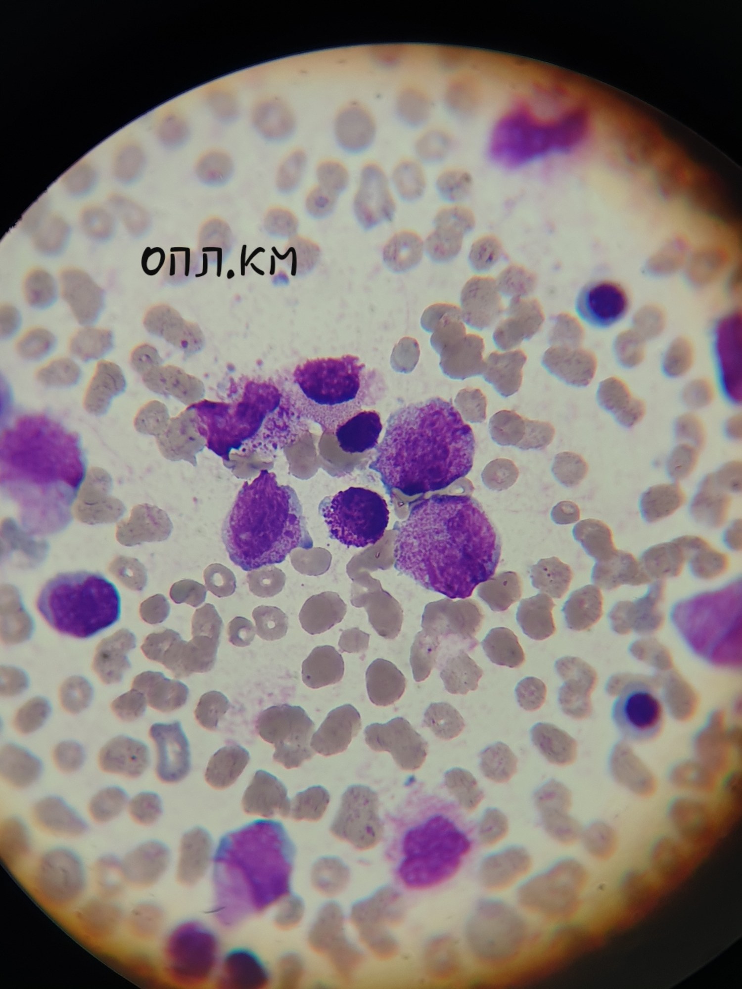
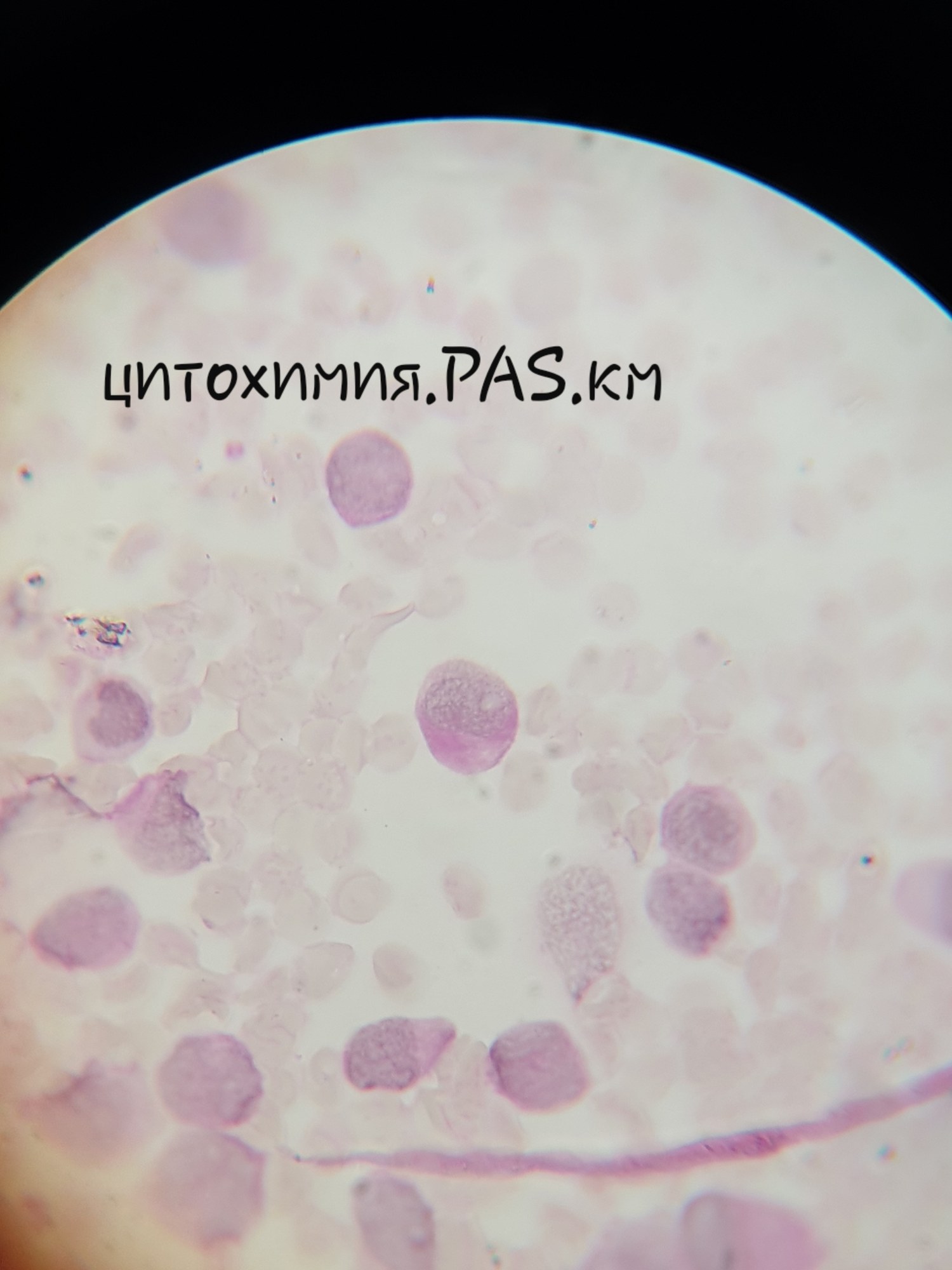
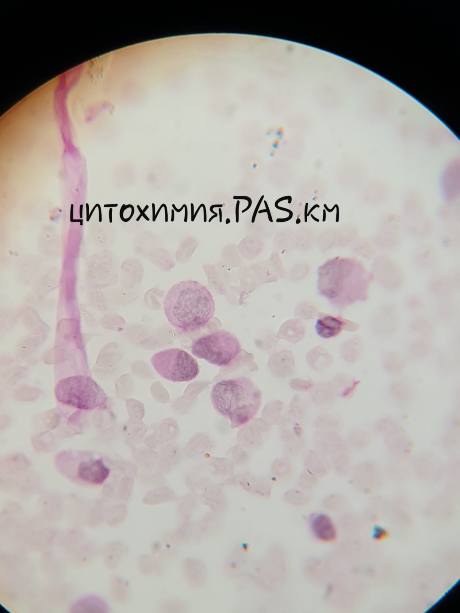
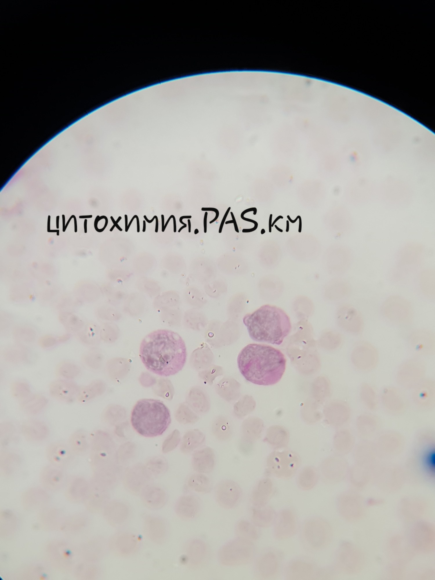
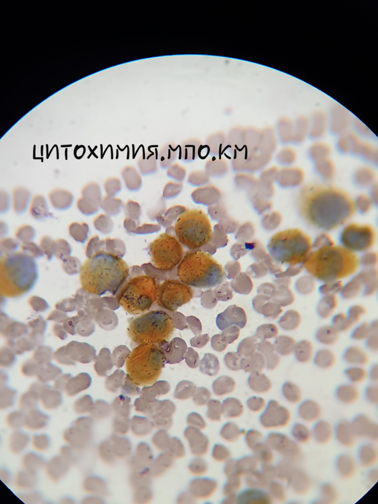
AML (M3) An 18-year-old female presented to the emergency department with intestinal bleeding and pancytopenia. Bone marrow was sent for consultation. Myelogram showed hypercellular bone marrow with an increased number of promyelocytes, including atypical promyelocytes. By the time the patient was transferred to our department, she had developed severe hemorrhagic syndrome. From 26.07 to 28.07, leukocytosis increased from 8.0 to 55.0. On 26.07, treatment according to the AIDA protocol was initiated. On 29.07, leukocytes were 45.0, erythrocytes 2.41, hemoglobin 86, platelets 40 (differential count: blasts 4%, promyelocytes 30%, myelocytes 27%, atypical promyelocytes 30%, segmented neutrophils 3%, lymphocytes 1%, monocytes 5%). Myelogram: 8.0% blasts, 37.0% atypical promyelocytes. Cytochemistry: MPO: strongly positive in 100% of blasts. PAS: positive in diffuse and diffuse-granular form in 88%. Immunophenotyping: CD117+CD33+CD13+CD15+MPO+Lysozyme+CD65+CD11b+CD45+ (consistent with AML) FISH: PML/RARa chimeric gene detected in 90% of cells. Blood smears and myelogram – Romanowsky stain. All photos 1000x
Acute Promyelocytic Leukemia (AML M3) Diagnosis
A clinical case of acute promyelocytic leukemia (AML M3) diagnosis, including blood and bone marrow cell morphology, cytochemistry (MPO, PAS), and PML/RARa translocation detection.