
🎓 Iron: When Excess Becomes Toxic
Iron is an essential trace element, vital for DNA synthesis and cellular metabolism. However, the human body lacks an active mechanism for eliminating its excess. When iron levels exceed physiological needs, it transforms into a dangerous oxidant.
Let’s explore modern approaches to the pathophysiology, diagnosis, and monitoring of iron overload based on current clinical guidelines. 🧬
🧪 Mechanisms of Damage With excessive iron accumulation, the transport protein transferrin becomes saturated. Free, unbound iron appears in the bloodstream – known as labile plasma iron.
This triggers a cascade of pathological reactions: Active oxygen species (free radicals) are formed. Lipid peroxidation of cell membranes is initiated. Cell organelles are destroyed, leading to subsequent tissue death (cytolysis).
🩸 Main Causes of Accumulation
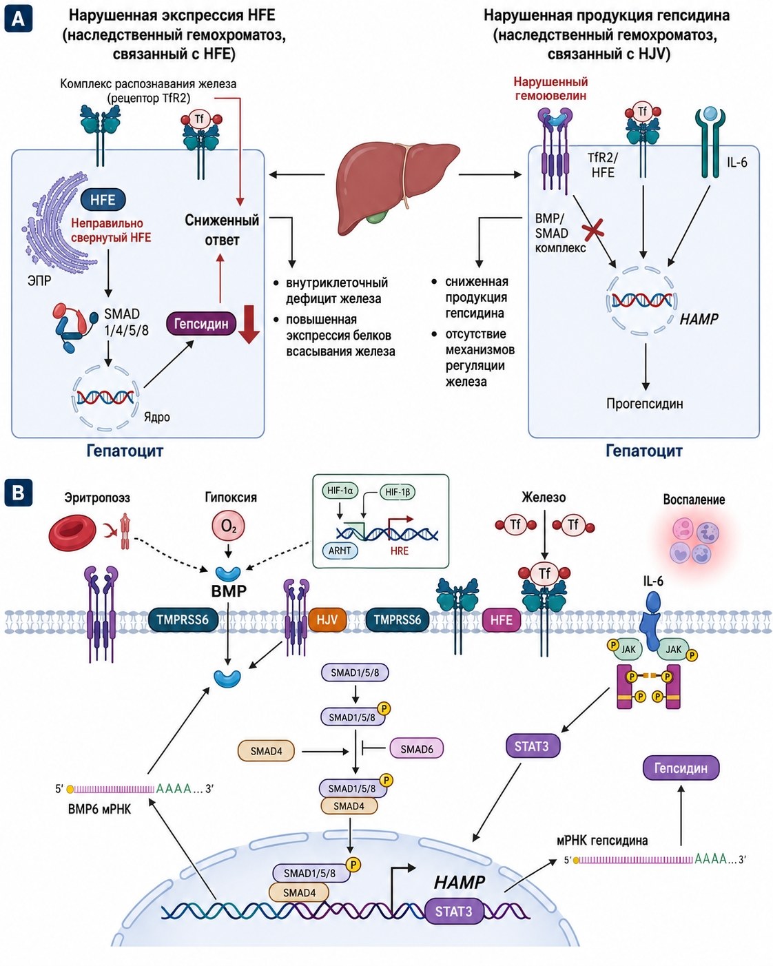
Hereditary Hemochromatosis: A genetically determined disorder where intestinal iron absorption from food significantly increases due to hepcidin deficiency.
Erythropoietic Hemosiderosis: Occurs in diseases with ineffective erythropoiesis (thalassemia, myelodysplastic syndromes).
Transfusional Overload: A consequence of multiple red blood cell transfusions, where each unit of blood introduces approximately 200 mg of iron into the body.
🏥 Clinical Targets Iron accumulates in parenchymal organs, leading to severe complications:
Liver: Fibrosis, cirrhosis, and a high risk of hepatocellular carcinoma. Heart: Development of specific cardiomyopathy and heart failure. Endocrine system: Damage to the pancreas (diabetes mellitus) and pituitary gland (impaired growth and reproductive function).
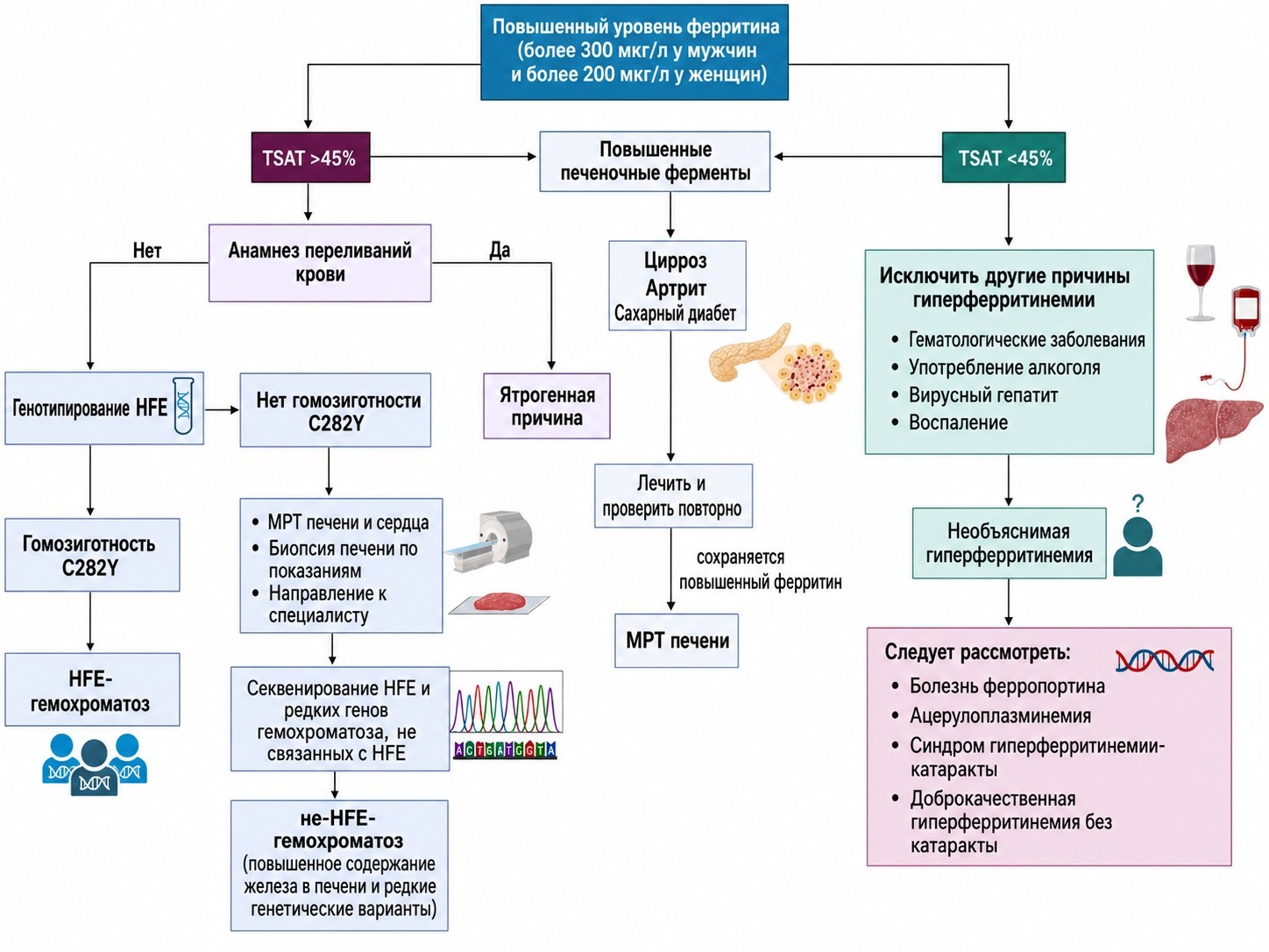
🔍 Modern Diagnostics Primary screening includes assessing two indicators: ✅ Serum Ferritin: Reflects total iron stores (levels above 200–300 µg/L require attention). ✅ Transferrin Saturation: A value above 45% is a marker of pathological accumulation.
The gold standard for assessing the severity of the condition today is T2-weighted magnetic resonance imaging (MRI). This non-invasive method allows for highly accurate measurement of iron concentration directly in liver and myocardial tissues, which is critically important for adjusting chelation therapy or prescribing therapeutic phlebotomy.
📈 Monitoring Patients with confirmed overload require regular monitoring of ferritin levels (every 3–6 months) and annual assessment of kidney function, thyroid gland, and blood glucose levels. Timely initiation of treatment can not only halt the progression of fibrosis but also reverse some toxic effects in the myocardium.
For citation: Chatzikalil E, Delaporta P, Bistas K, Kattamis A. Iron Overload: Pathophysiology, Diagnosis and Monitoring. International Journal of Laboratory Hematology. 2026;0:1–10.


