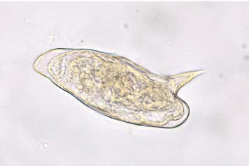
🔬Schistosoma mansoni egg. Note the prominent lateral spine (400×). ✅Schistosoma mansoni is found in Africa, especially in tropical areas and the Nile Delta, southern Africa and Madagascar, Brazil, Venezuela, Suriname, and some Caribbean islands, including Puerto Rico. Adult S. mansoni live primarily in the portal vein and inferior mesenteric vein. ✅Initial egg deposition in the large intestine can cause abdominal pain and dysentery with copious blood and mucus in the stool. At this time, eggs may be found in the feces. Chronic infection can lead to hepatic fibrosis and portal hypertension depending on the number of worms present; at this stage, it may be more difficult to find eggs in the feces. ✅Eggs measure 116 to 180 µm by 45-58 µm, are oval, and possess a large, distinct lateral spine that protrudes from the side of the egg near one end (photograph below). Miracidial movement within the egg may be evident in unfixed material and, if seen, indicates an active infection. ✅Concentration methods (Parasep method) may be required to detect eggs, as individuals with chronic infection may have a small number of eggs. Source: McPherson - Henry’s Clinical Diagnosis and Management by Laboratory Methods, 24ed 2022
Schistosomiasis Diagnosis: Schistosoma mansoni Eggs in Stool
Microscopy features of Schistosoma mansoni eggs: prominent lateral spine, size, manifestations of intestinal schistosomiasis, and egg detection using the Parasep method.