

⚠ Brugia - microfilaria. Microscopy of a wet blood smear 🩸
To catch this parasite, blood is collected at night from 10 PM to 2 AM. Microfilariae are visible in a direct wet smear. The species can be differentiated in a smear stained with Romanowsky-Giemsa (swipe through the carousel for the algorithm, in English).
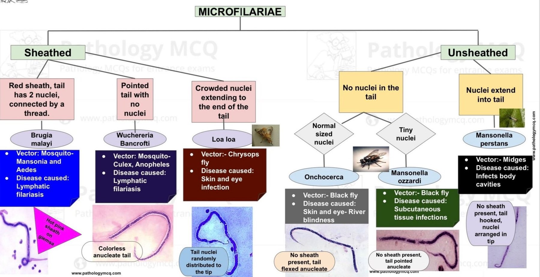
🐍 Algorithm for differential diagnosis of microfilariae species:
❗Sheathed: ✅ Brugia malayi Red sheath, 2 nuclei on the tail connected by a thread. Vectors: Mansonia and Aedes mosquitoes. Disease: lymphatic filariasis. Bright pink sheath when stained with Giemsa.
✅ Wuchereria bancrofti Pointed tail without nuclei. Vectors: Culex and Anopheles mosquitoes. Disease: lymphatic filariasis. Colorless tail without nuclei.
❗Unsheathed: ✅ Loa loa Dense nuclei extending to the end of the tail. Vectors: deer fly (Chrysops). Disease: cutaneous and ocular infections. Tail nuclei distributed to the very tip.
✅ Onchocerca Normal-sized nuclei. Vectors: black fly. Disease: cutaneous and ocular infections — river blindness. Sheath absent, tail curved, without nuclei.
✅ Mansonella ozzardi Very small nuclei. Vectors: black fly. Disease: subcutaneous tissue infections. Sheath absent, tail pointed, without nuclei.
✅ Mansonella perstans Nuclei extend into the tail. Vectors: midges. Disease: body cavity involvement. Sheath absent, tail curved, nuclei located at the tip.


