

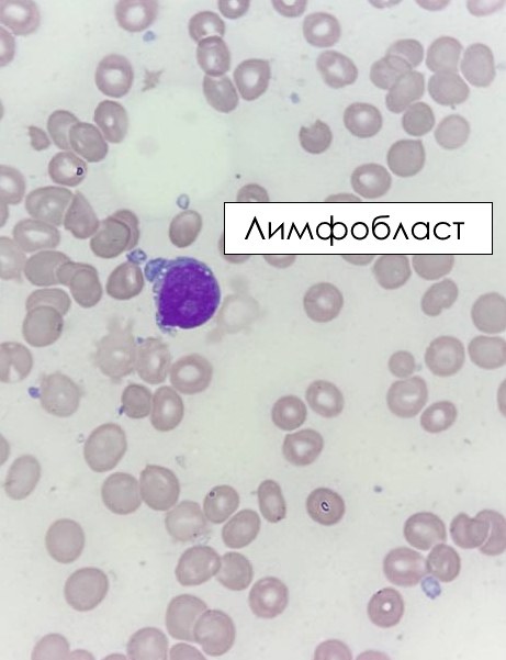
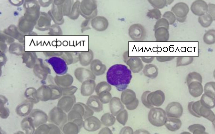
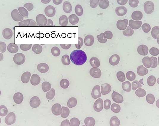
T-lymphoblasts or T-prolymphocytes? A clinical case. A 63-year-old female. Medical history. One year ago, she was diagnosed with T-prolymphocytic leukemia, with leukocytosis in CBC: 267 x 103/µL. Several courses of chemotherapy were administered according to the venetoclax+bendamustine regimen. ❗CBC today: ↓↓↓ White Blood Cells (WBC) 0.16 x 10³/µL!! (Normal 3.70 - 9.50) ↓ Red Blood Cells (RBC) 2.99 x 10⁶/µL (Normal 4.00 - 5.20) ↓ Hemoglobin (HGB) 85 g/L (Normal 118 - 150) ↓ Platelets (PLT) 88 x 103/µL (Normal 150 - 400) 🔬Microscopy of blasts in the images below from a smear of hypocellular bone marrow aspirate. Do not confuse them with T-prolymphocytes – there are similar features: cytoplasmic blebbing (cytoplasmic protrusions), a pale nucleolus. It is important to look at chromatin dispersion here! 💡 Immunophenotyping. Conclusion. The bone marrow is poor in cellular events. In the examined sample, the mononuclear cell polygon constitutes 81.65%. It is dominated by cells with the immunophenotype CD45+CD2+CD7+CD5+CD4-CD8+CD3+CD1a-CD56-CD38+HLA-DR-+CD3cyt+, which corresponds to mature T-ALL, T-IV variant.
❗Morphology description from the WHO 2022 classification of hematolymphoid tumors: “Lymphoblasts in T-ALL are morphologically indistinguishable from those in B-ALL. In blood/BM smears, blasts have scanty basophilic agranular or rarely vacuolated cytoplasm, round, irregular, or convoluted nuclei. They vary from small to medium-sized with a high nuclear-to-cytoplasmic ratio with condensed chromatin and no apparent nucleoli, to larger blasts with finely dispersed chromatin and relatively prominent nucleoli. Occasionally, blasts in T-ALL may resemble more mature lymphocytes, and then immunophenotypic studies are required to distinguish this disease from mature (peripheral) T-cell leukemia/lymphoma.”


