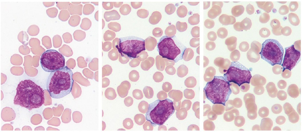
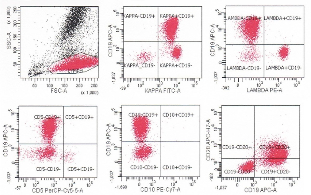
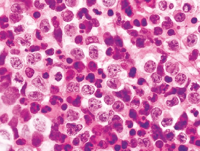
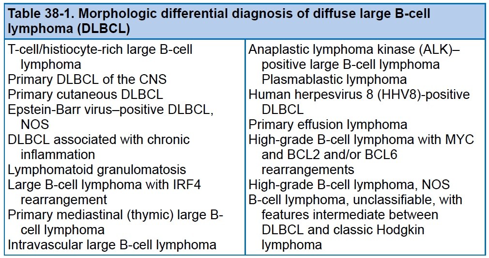
🔬A case of relapsed diffuse large B-cell lymphoma (DLBCL) with peripheral blood involvement in a 59-year-old woman. ✅PHOTO 1: peripheral blood microscopy. 🩸Leukocytosis 69.1 × 10^9/L with 57% atypical lymphocytes. ✅PHOTO 2: IFT of this patient Tumor lymphocytes are positive for CD19 and CD20, negative for CD5 and CD10, and demonstrate monotypic staining of surface immunoglobulin kappa light chain. ✅DLBCL represents a biologically diverse diagnostic category that includes large B-cell neoplasms with a diffuse growth pattern and the absence of diagnostic criteria for other lymphomas. DLBCL can arise de novo or as a result of transformation from a low-grade B-cell process; a well-known example of the latter is Richter’s transformation of chronic lymphocytic leukemia/small lymphocytic lymphoma into DLBCL. Risk factors for DLBCL include immunodeficiency and advanced age. DLBCL can have a prominent extranodal component; up to 40% of cases are initially confined to extranodal sites. The most common extranodal site is the gastrointestinal tract; others include the spleen, bones, testes, thyroid, and liver. B symptoms may or may not be present. Peripheral blood involvement in DLBCL is rare; an example case is shown in photos 1 and 2. Lymph nodes with DLBCL show effaced normal architecture, replaced by diffuse sheets of large lymphoid cells. There are three common morphological variants: the centroblastic variant is the most common and consists of medium to large cells with scant basophilic or amphophilic cytoplasm, round nuclear contours, fine vesicular chromatin, and multiple small membrane-bound nucleoli (see photo 3). ✅PHOTO 3: Retroperitoneal lymph node, core needle biopsy (H&E stain - hematoxylin and eosin). Shows centroblastic morphology with scant cytoplasm, round to oval nuclei, and multiple membrane-bound nucleoli. ✅PHOTO 4: Morphological differential diagnosis of diffuse large B-cell lymphoma 👉Source: D. Dorfman - Color Atlas of Flow Cytometry, 2023
Diffuse Large B-Cell Lymphoma: A Case of Leukemization
A clinical case of DLBCL leukemization: morphology of atypical lymphocytes in blood, immunophenotyping by flow cytometry, and lymph node biopsy.