

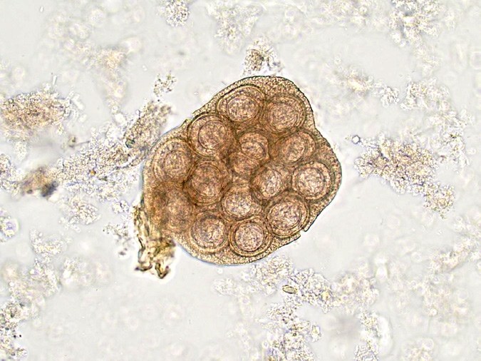
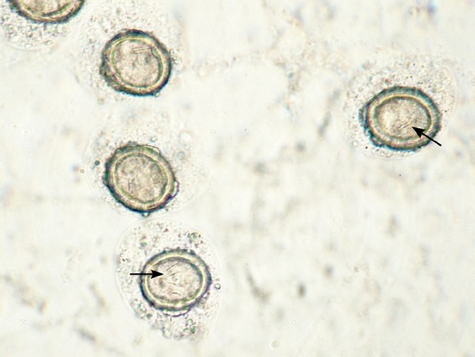
Dipylidium caninum (cucumber tapeworm) eggs are often found in an egg capsule measuring ~100–200 µm, containing 5–30 eggs (photo 1). Individual eggs are round, approximately 30–50 µm in diameter, and have a thick, radially striated embryophore containing an embryo with three pairs of hooks (photo 2). Clinical Aspects
- Commonly found in dogs and cats, but incidence is decreasing.
- Eggs are not consistently shed and may not be detected in feces.
- Clinical disease is rare. Perianal irritation is most common.
Prognosis is excellent with therapy, including control of intermediate hosts (e.g., fleas). ✅ Dipylidium caninum is a tapeworm that can parasitize the human intestine without causing any symptoms. The photos show microscopy of dog feces. Dipylidium caninum ✅ Photo №1. Eggs in capsule, dog, 20× objective; ✅ Photo №2 Individual eggs. Note the individual eggs with small hooks (arrows in photo 2), 100× objective.
👉 Source: Andrew G. Burton - Clinical Atlas Cytology and Hematology 2nd Edition, 2024 download a collection of books on laboratory veterinary medicine here →


