

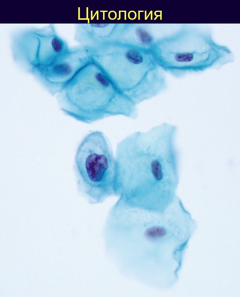
Cervical cytology. Dyskeratotic nuclei are characterized, as in this background cell, by chromatin clumping, hyperchromasia, and an irregularly thickened nuclear membrane.
This is a characteristic cytological sign of neoplastic changes in squamous epithelium associated with HPV infection.
Note: The diagnosis is based precisely on nuclear abnormalities — without them, the term “dyskeratosis” is not applied.
Source: M. Sheaff, N. Singh. Cytopathology: An Introduction (2013).
- —
Hyperchromasia is a term used by pathologists to describe a cell nucleus that appears significantly darker than normal under microscopic examination. This occurs because a hyperchromatic nucleus absorbs more stain (typically hematoxylin and eosin) compared to a normal nucleus, giving it a more intense blue or purple color. 💎 Find more useful cytology materials in our LAB-TESTS library. The link with access to the library is automatically sent to you via private message after paying for a VK donut Labformula subscription.


