

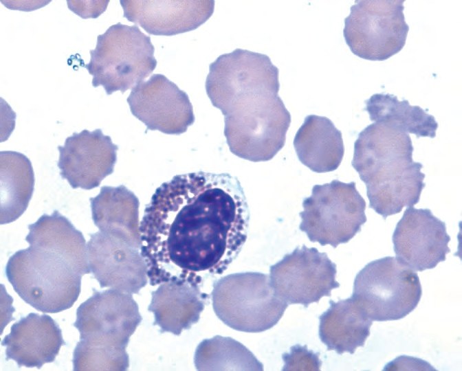
Eosinophil dysplasia in a peripheral blood smear.
The nucleus is irregularly oval, unsegmented, pseudo-Pelger type, with markedly condensed chromatin.
The agranular part of the cytoplasm retains weak basophilia, indicating impaired maturation and retention of ribosomes.
Granules are unevenly distributed, varying in size and staining characteristics. ⭐ Source: Giuseppe d’Onofrio - Morphology of Blood Disorders 2015


