

🔬Direct microscopic examination of stool: the red arrow shows the budding of Candida.
👉Source: Hameed, Ali & Ali, Sabah & Ahmed, Luma. (2018). The Prevalence of Candida spp. Among Children with Diarrhea in Baqubah-Iraq. 1. 34-43.
Morphology of Candida yeast-like fungi in a native stool smear during coprological examination: detection of budding cells under the microscope.
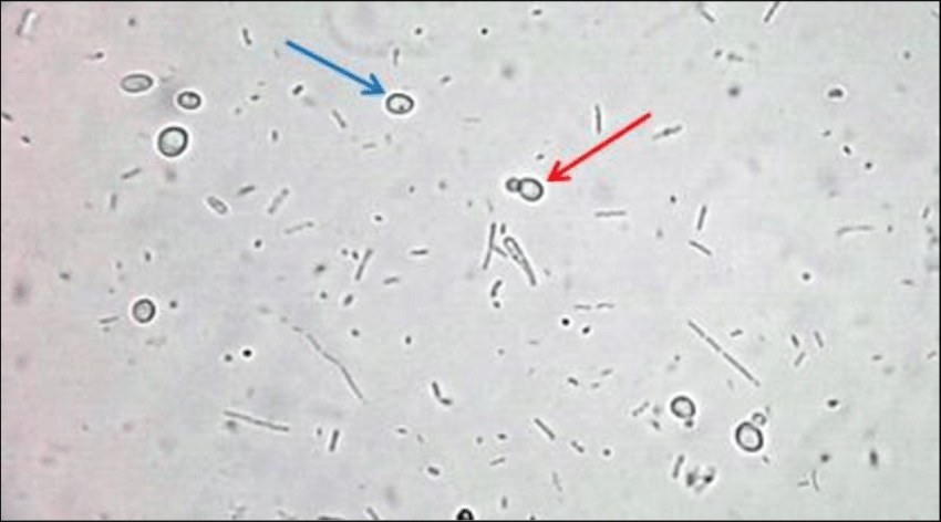
🔬Direct microscopic examination of stool: the red arrow shows the budding of Candida.
👉Source: Hameed, Ali & Ali, Sabah & Ahmed, Luma. (2018). The Prevalence of Candida spp. Among Children with Diarrhea in Baqubah-Iraq. 1. 34-43.

Morphology of yeast-like fungi in stool during coproscopy. Microscopy of preparations with Lugol's solution and methylene blue at ×400 magnification.

Microscopic examination of stool during coprogram: detection and morphology of budding yeast-like fungal cells stained with methylene blue.

Detection of mycelial filaments and budding yeast cells in urine sediment: microscopic picture in mycosis and contamination with vaginal discharge.