

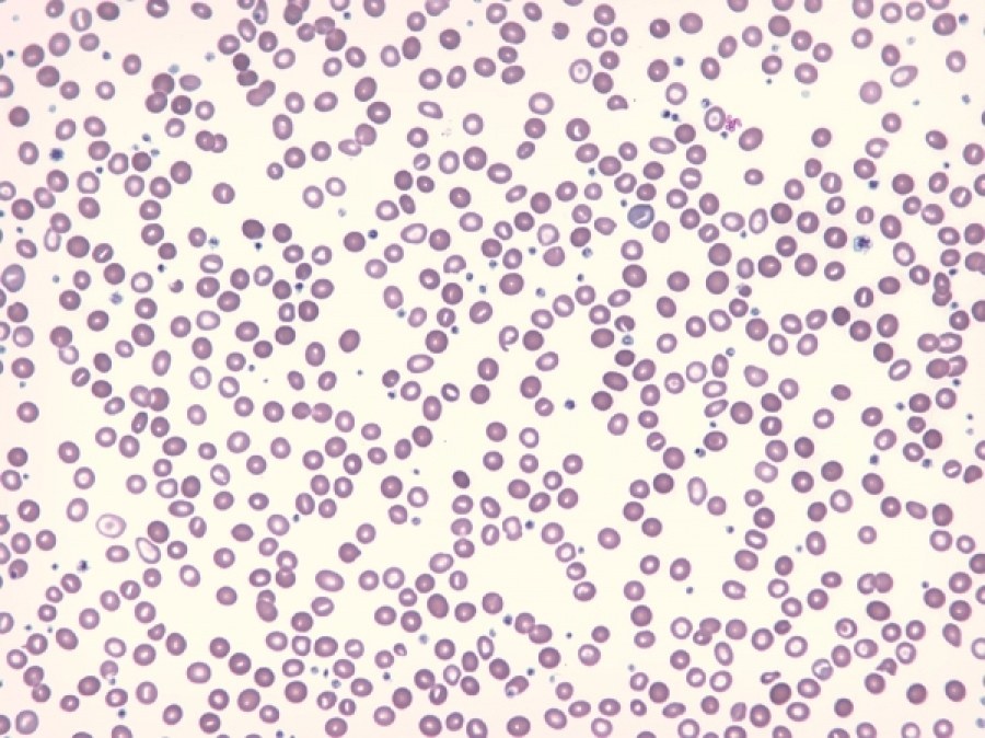
Anisochromia due to a dual red blood cell population: one well-hemoglobinized, with a spherocytic appearance (transfused red blood cells), and the other – microcytic and hypochromic red blood cells.
Clinical information: Iron deficiency anemia, blood transfusion.
A 75-year-old woman with a history of iron deficiency anemia treated with iron supplements. Referred to the emergency department with symptomatic microcytic iron deficiency anemia. 3 units of packed red blood cells were transfused.
Biological information: Hemogram before transfusion: Hb 56 g/L, MCV 63.3 fL, MCH 14.7 pg; MCHC 239 g/L; RDW 22.4%, leukocytes and platelets within normal limits. Iron metabolism: serum iron: 14 µg/dL, Ferritin: <6 ng/mL.
Hemogram after transfusion (image): Hb 97 g/L, Hematocrit 37.2, MCV 77.3 fL, MCH 20.2 pg, MCHC 265 g/L, RDW 28.6%. Leukocytes and platelets within normal limits.
Sample type: Peripheral blood
Staining type: Wright
Author(s): María Pilar Ricard Andrés
Workplace: Hospital Universitario Fundación Alcorcón


