

Sometimes endothelial cells in an image look very similar to hairy cell leukemia cells, but the chromatin structure is significantly different.
🔬 Source: from the archive of Brazilian hematology specialist Marcio Melo
Morphological differences between endothelial cells and hairy cell leukemia cells in peripheral blood smear: chromatin and cytoplasm features.
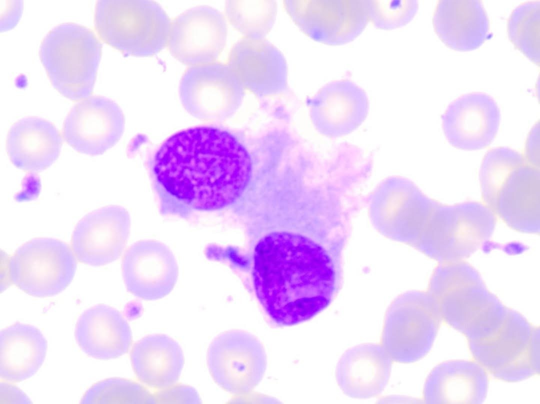
Sometimes endothelial cells in an image look very similar to hairy cell leukemia cells, but the chromatin structure is significantly different.
🔬 Source: from the archive of Brazilian hematology specialist Marcio Melo

Morphological features of monocytes in peripheral blood smear: variability of nuclear shape, differential diagnosis with other leukocytes.

Criteria for monocyte identification in blood smears: morphological differences from lymphocytes, reactive forms, and neutrophils during differential leukocyte count.

Evaluating lymphocyte morphology in blood smears: vacuolization, cytoplasmic villi, and differential diagnosis with oncohematological diseases.