

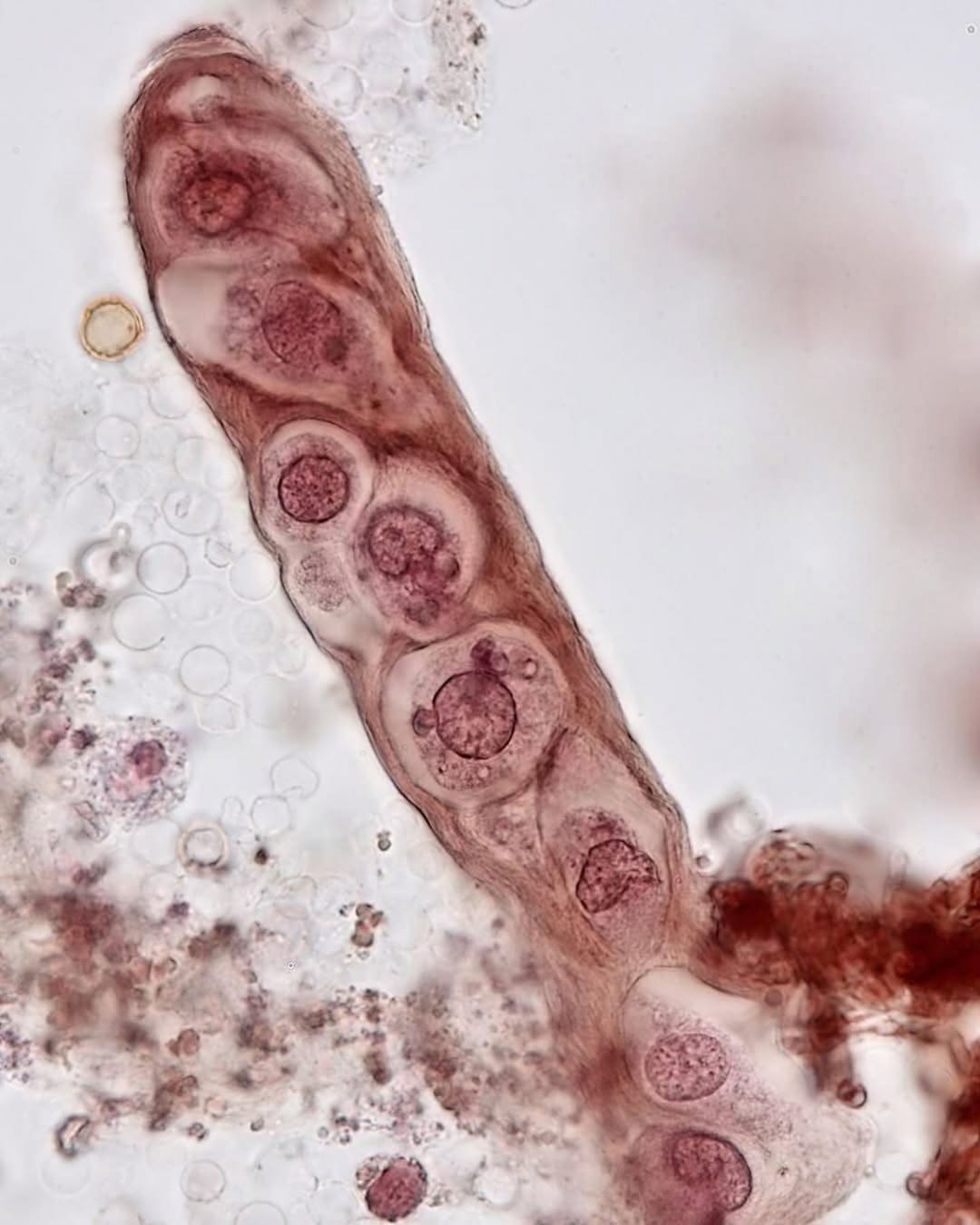
🔬EPITHELIAL CASTS are microscopic structures that form within the renal tubules as a result of the adhesion of epithelial cells detached from the tubular epithelium. These cells can gather into disorganized clumps or form organized layers, characterized by large nuclei and low cytoplasmic content. The appearance of such casts is especially characteristic of the diuresis recovery phase after acute renal failure caused by tubular necrosis. Their detection in urine sediment is a clinically significant observation, as it is usually associated with pathological conditions.
Their presence is associated with: 🔘 Acute tubular necrosis 🔘 Toxin intake (mercury, diethylene glycol, salicylates, etc.) 🔘 Inflammatory kidney diseases
💎 Find more useful materials on urine examination in our LAB-TESTS library. The link with access to the library is automatically sent to you via private message after paying for a VK donut Labformula subscription.


