

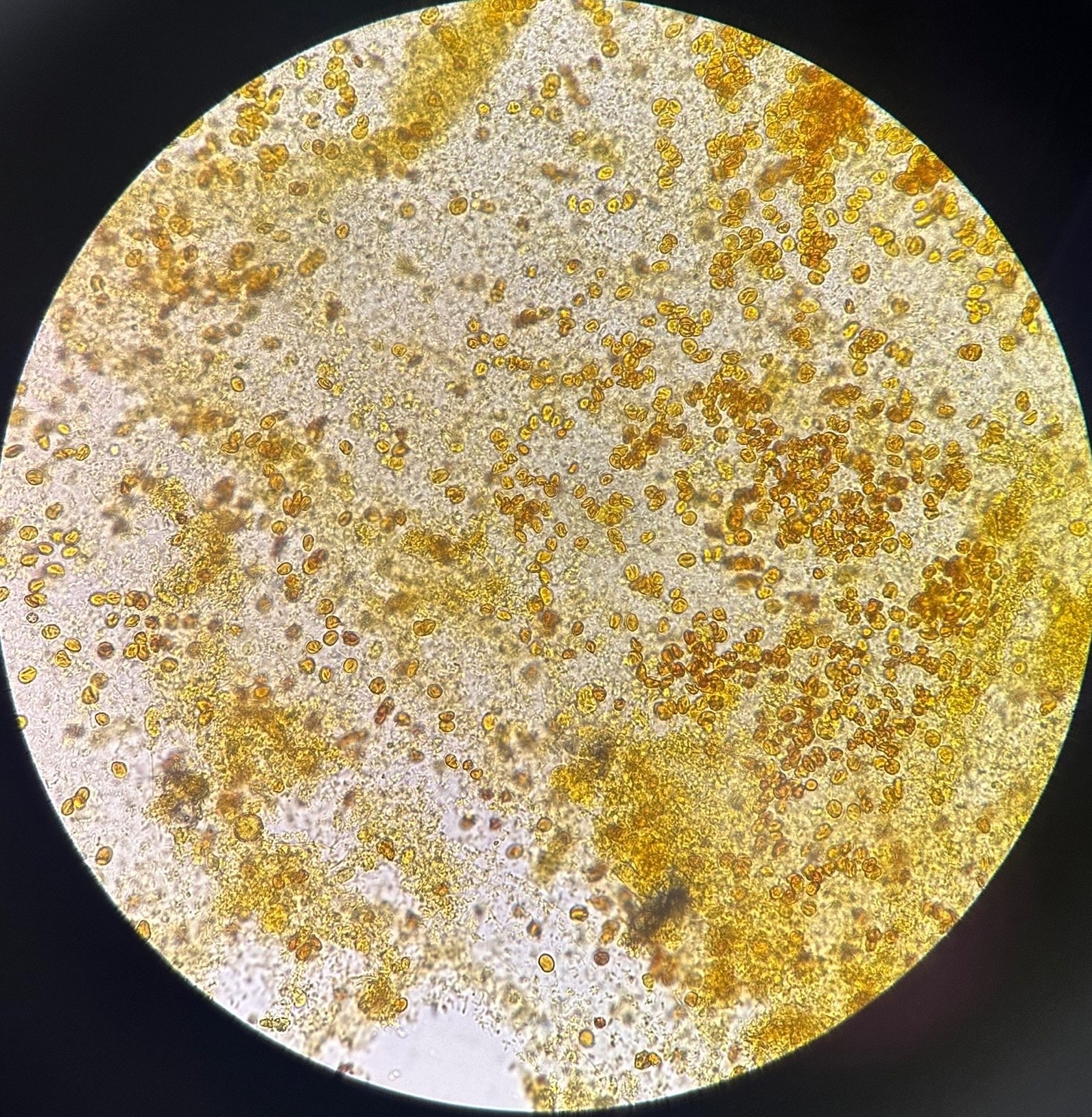
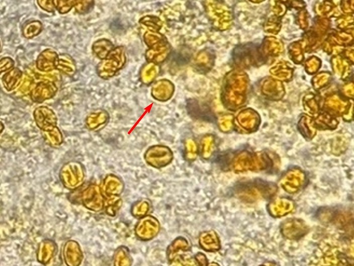
Erythrocytes in stool. Lugol’s solution preparation. 🙏 Images kindly provided by clinical laboratory diagnostics physician, Anna Ivanova, Moscow.
REFERENCE INFORMATION In stool, erythrocytes lose their smoothness due to:
- Enzymes and acids - pepsin, hydrochloric acid (in gastric bleeding) - destroy the membrane.
- Mechanical damage due to peristalsis.
- Changes in pH and osmolarity in the intestine - cells shrink or swell.
Result: erythrocytes under the microscope appear deformed, fragmented, or colorless (leached). However, intact erythrocytes can also be found (one of them is indicated by an arrow in the second photo).


