

The Splendore-Hoeppli phenomenon is a rare occurrence in the diagnosis of infections.
The Splendore-Hoeppli phenomenon (SHP) is a rare phenomenon, first described in 1908. It manifests as an accumulation of eosinophilic material around bacteria (Actinomyces, Nocardia), fungi (Aspergillus, Candida), parasites (Schistosoma), or foreign bodies (suture material). SHP is usually associated with chronic inflammatory processes and tissue necrosis.
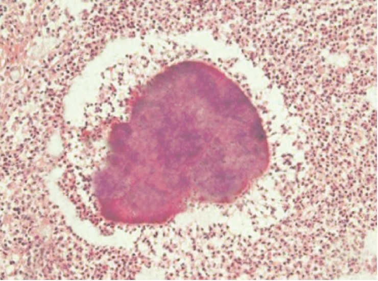
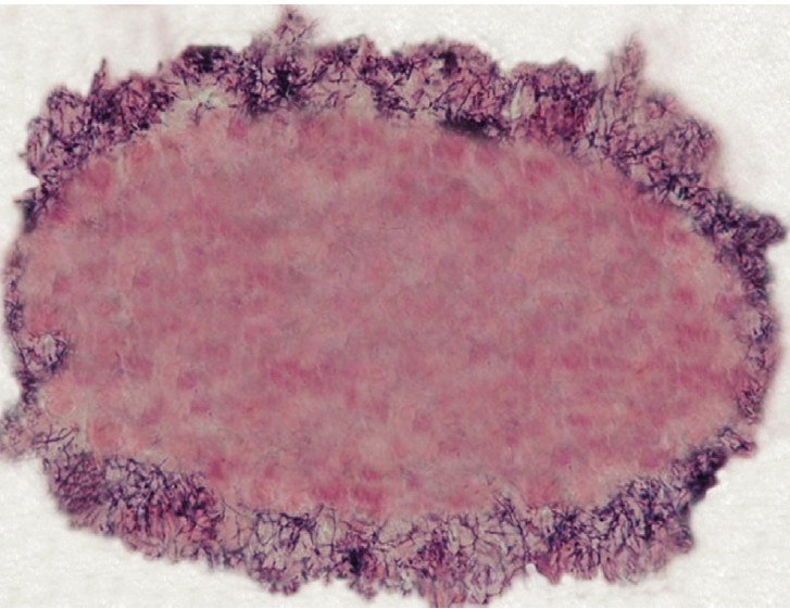
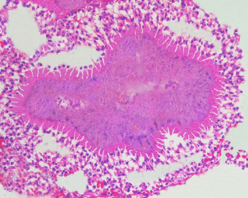
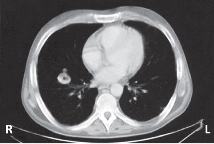
In 2020, a case of pulmonary actinomycosis with “sulfur granules” and the SHP phenomenon in a 67-year-old patient was described in the journal Diagnostic Cytopathology. Diagnosis included lung CT and transbronchial aspiration. Gram staining revealed gram-positive bacilli, and histology confirmed the SHP phenomenon.
🔬 Important to remember: if you see this phenomenon, it is necessary to suspect an infection, such as actinomycosis, and perform specific stains.
Figure 1: Image of a “sulfur granule” surrounded by inflammatory cells, Papanicolaou stain, ×200 magnification.
Figure 2: Cell block from transbronchial needle aspiration, demonstrating a “sulfur granule” with the Splendore-Hoeppli phenomenon (H×E stain, ×200 magnification).
Figure 3: Cell block with numerous gram-positive bacilli located at the periphery of an oval mass (Gram stain, ×400 magnification).
Figure 4: Chest computed tomography showing an irregular mass with central cavitation in the right lung (Pulmonary actinomycosis).
💎 Find even more useful materials on cytology in our LAB-TESTS library. The link with access to the library is automatically sent to you via private message after paying for a VK donut Labformula subscription.
Source: Martínez-Girón R., Pantanowitz L. " Splendore-Hoeppli" phenomenon //Diagnostic cytopathology. – 2020. – Т. 48. – №. 12. – С. 1316-1317.


