

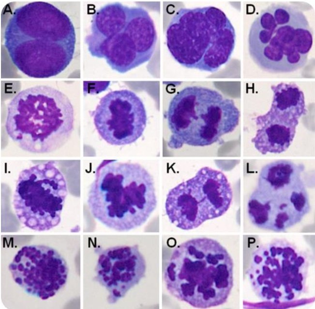
Clinical case: mitotic figures in myelodysplastic syndrome.
A 64-year-old man was admitted with progressive asthenia and pancytopenia. Complete blood count showed hemoglobin 7.5 g/dL, white blood cell count 1.2 × 10⁹/L, and platelet count 25 × 109/L. Bone marrow aspirate revealed 7% myeloblasts and 69% erythroblasts with dysplastic features: cytoplasmic vacuoles, nuclear budding, and multinucleated cells (panels A-D). Mitotic figures were observed in all phases of division in the erythroid lineage, including normal mitotic figures (panels E-H) and abnormal ones (panels I-L). Additionally, some erythroblasts presented with multiple micronuclei (panels M-P). Cytogenetic analysis revealed an abnormal clone: 43∼45,XY,-5,-7,-18,?add(19)(q13.3),+mar1,min[cp20]. The diagnosis of myelodysplastic syndrome (MDS) with excess blasts was confirmed.
Micronuclei/micronucleations are nuclear fragments that do not contain the complete cell genome, usually formed as a result of mitotic division errors or nuclear fragmentation during apoptosis. Micronuclei are a rare phenomenon in MDS and are associated with mitotic dysfunction. Recently, an oncogene-suppressing mechanism based on the phenomenon of mitotic catastrophe was described. This phenomenon leads to the death or senescence of mitotically abnormal and genomically unstable cells. The inability to activate this mechanism leads to persistent genomic instability and aneuploidy.
The question arises: does mitotic catastrophe participate in the physiopathology of MDS? A certain connection can be identified based on the mitotic figures in this clinical case, but this remains a topic for future research.


