

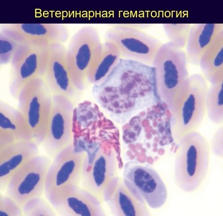
🦩 Blood of a greater flamingo (Phoenicopterus ruber).
The image shows:
- Heterophil (left) – with a pale basophilic nucleus and brick-red, spindle-shaped cytoplasmic granules.
- Eosinophil (right) – with a segmented basophilic nucleus and round/oval, brightly eosinophilic granules of various sizes.
- Monocyte (top) – with a constricted nucleus (reticular chromatin) and agranular basophilic cytoplasm.
Erythrocytes are also visible. Note that the cell shape is distorted due to adjacent elements. (Stain: Modified Wright’s method).
Source: Phillip Clark – Atlas of Clinical Avian Hematology, 2009. 🏛 Library “Veterinary Laboratory Medicine” ➡️ Level III subscription Level III - “Metamyelocyte” provides access to this book and the entire training course “Hematology of Exotic Animals”: Lesson 1: Reptile Blood Morphology 🦎 Lesson 2: Avian Blood Parasites 🐦 Lesson 3: Blood Features of Domestic Exotic Pets 🐷 Lesson 4: Differences Between Monkey and Human Blood 🐵
🔗 Details and subscription → vk.com/@leukoformula-3-level


