

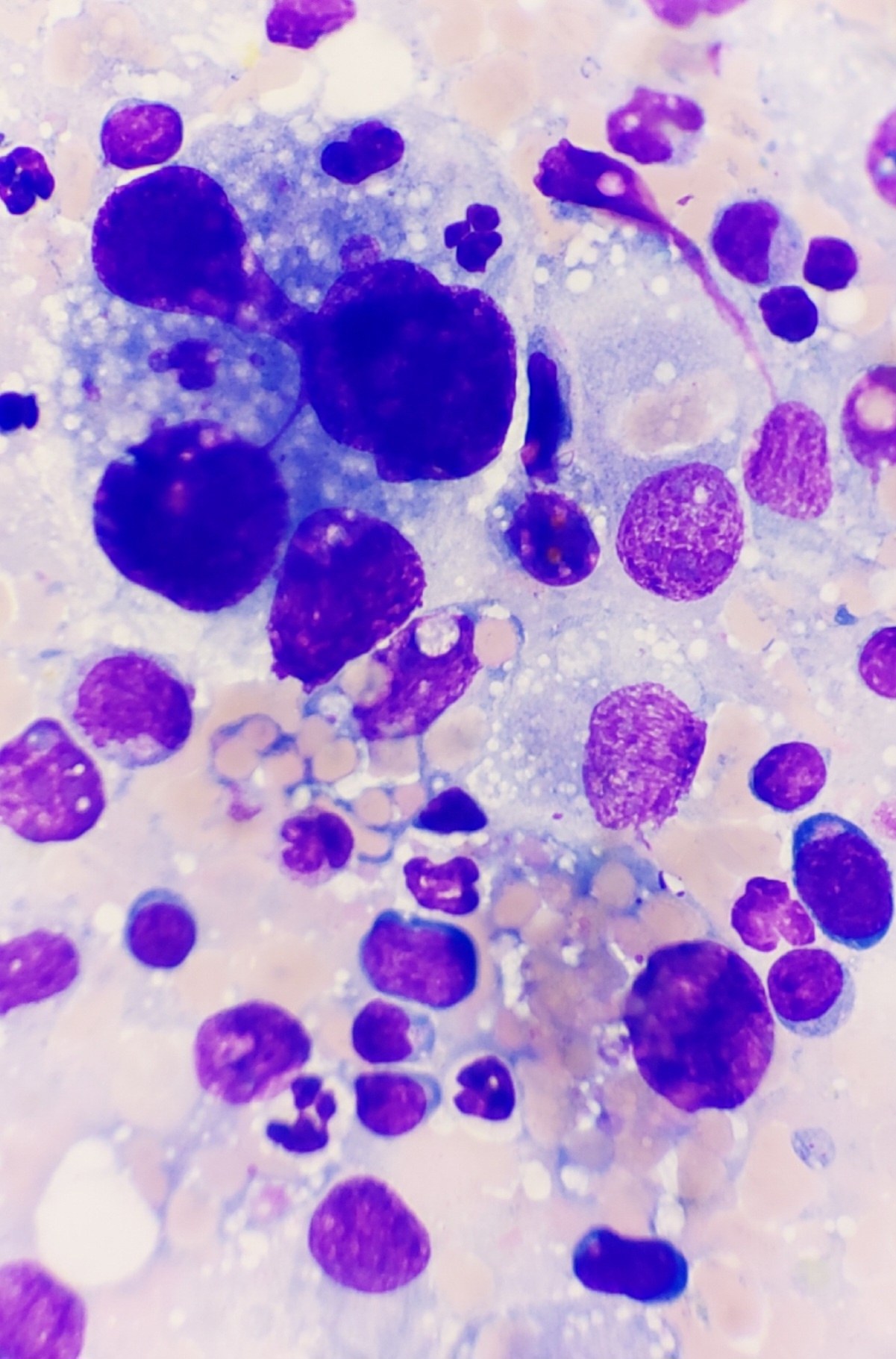
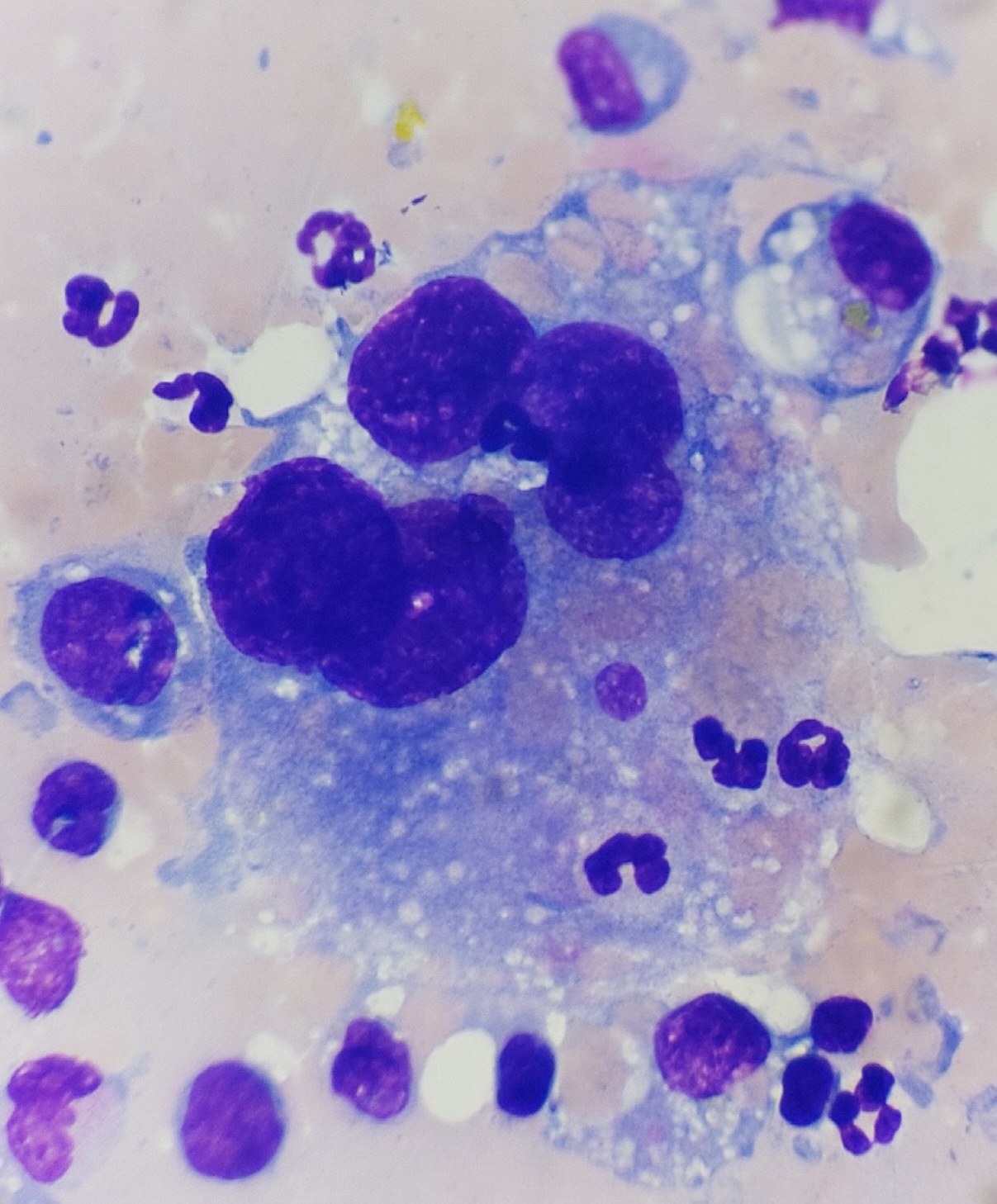
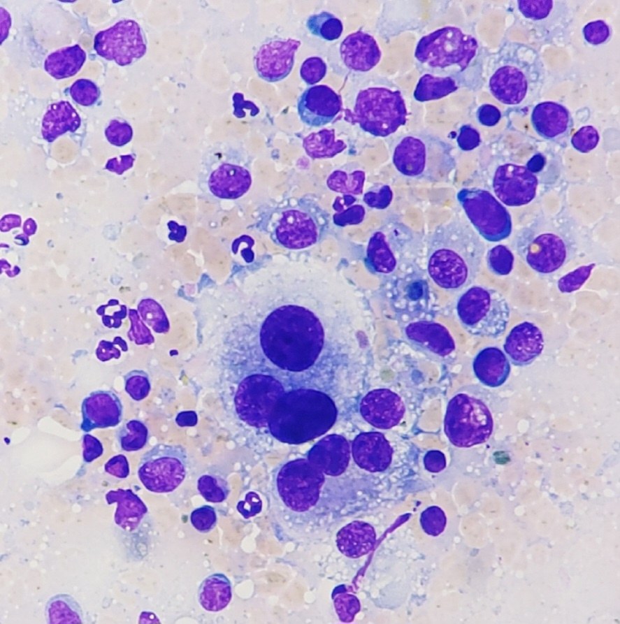
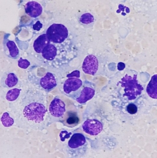
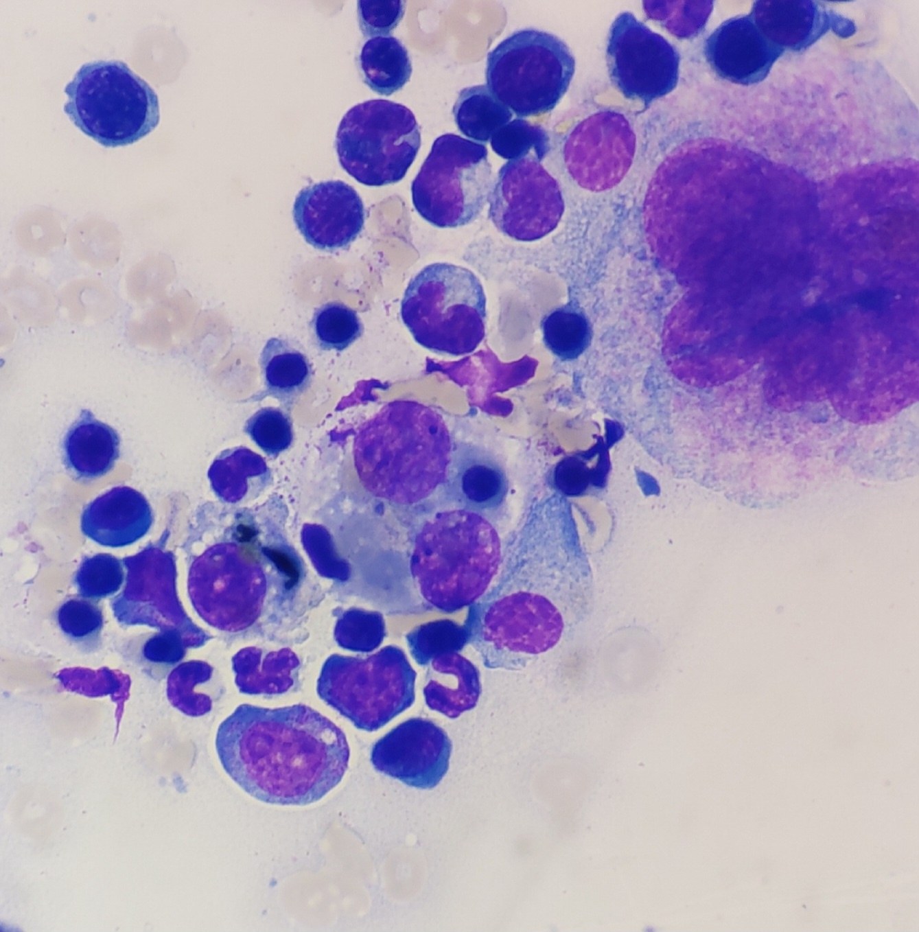
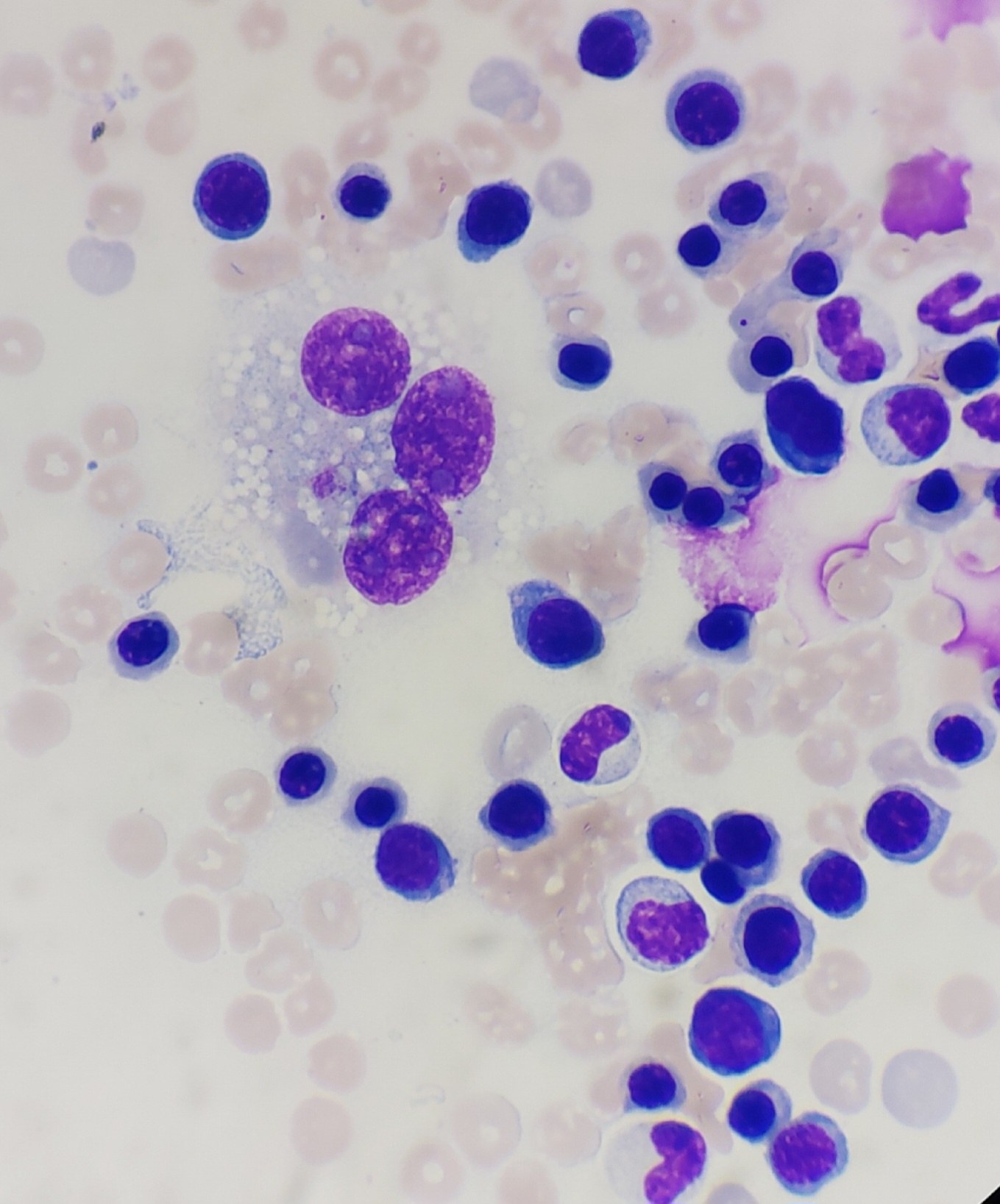
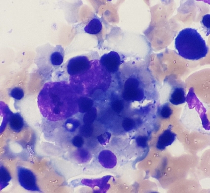
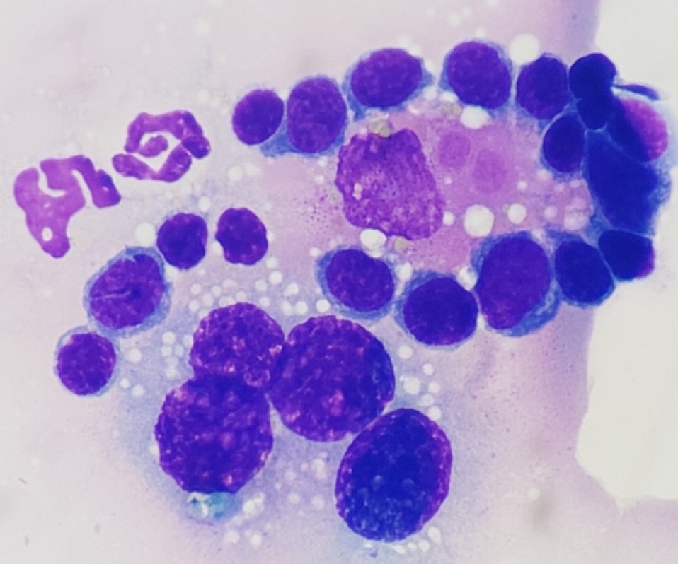
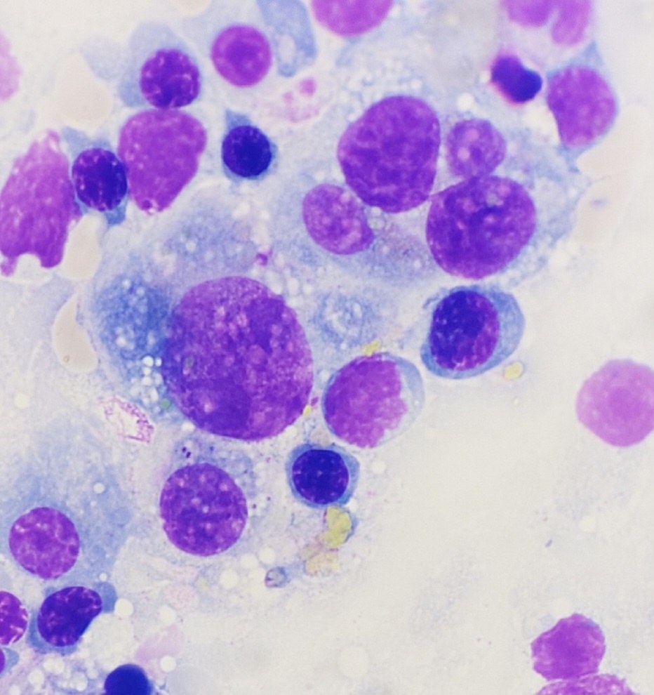
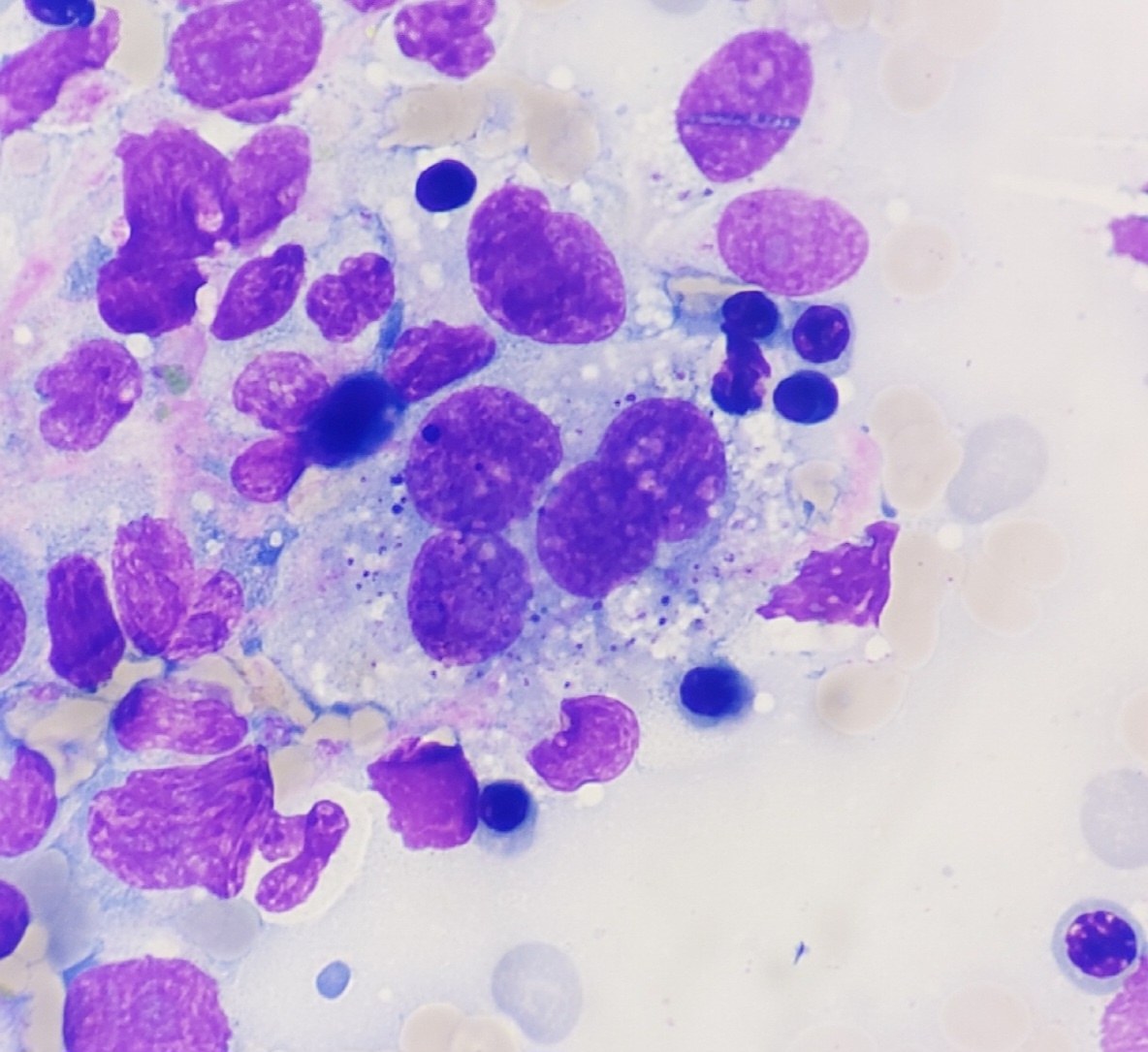
🔬 Hemophagocytic Histiocytic Sarcoma in a Dog: Lymph Node and Bone Marrow Cytology 📍 Clinical Case: Mixed-breed dog, female, 7 years old. Moderate regenerative anemia and moderate thrombocytopenia. 🧪 Cytology: First 4 photos — mesenteric lymph node aspirate. Next 6 photos — bone marrow biopsy. 🧫 Conclusion: 📌 Hemophagocytic histiocytic sarcoma in both locations. 📌 Bone marrow: marked erythroid hyperplasia (ineffective) and megakaryocytic hyperplasia. 📌 Thrombocytopenia of non-central origin.
🔍 Other features: Blood smear examination revealed isolated macrophages with signs of erythrophagocytosis. ❗ No blood parasite infections detected.
🙏 Case kindly provided by - Olga Belozerova, clinical pathologist, head of the laboratory at the veterinary clinic “Noah’s Ark”, Kirov.


