

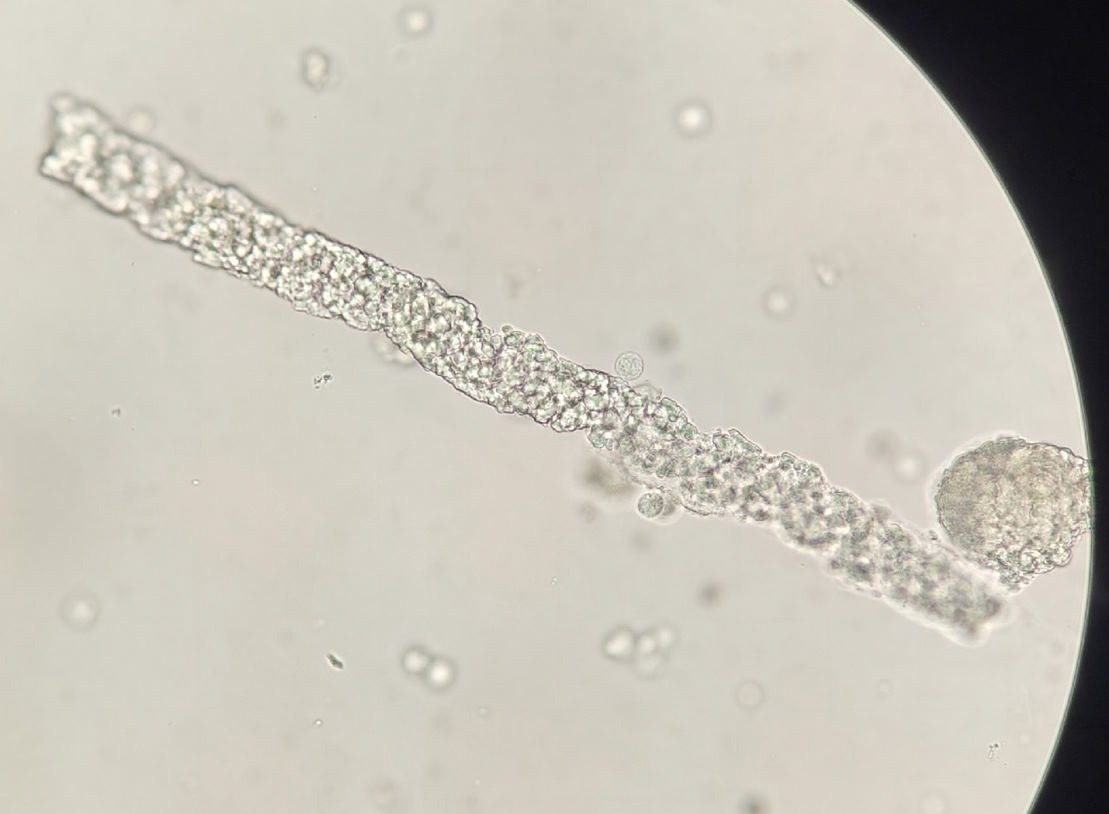
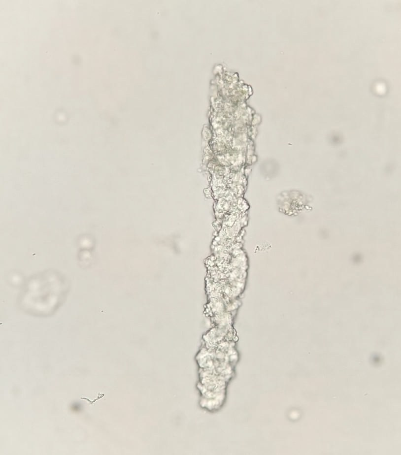
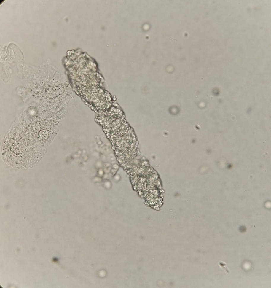
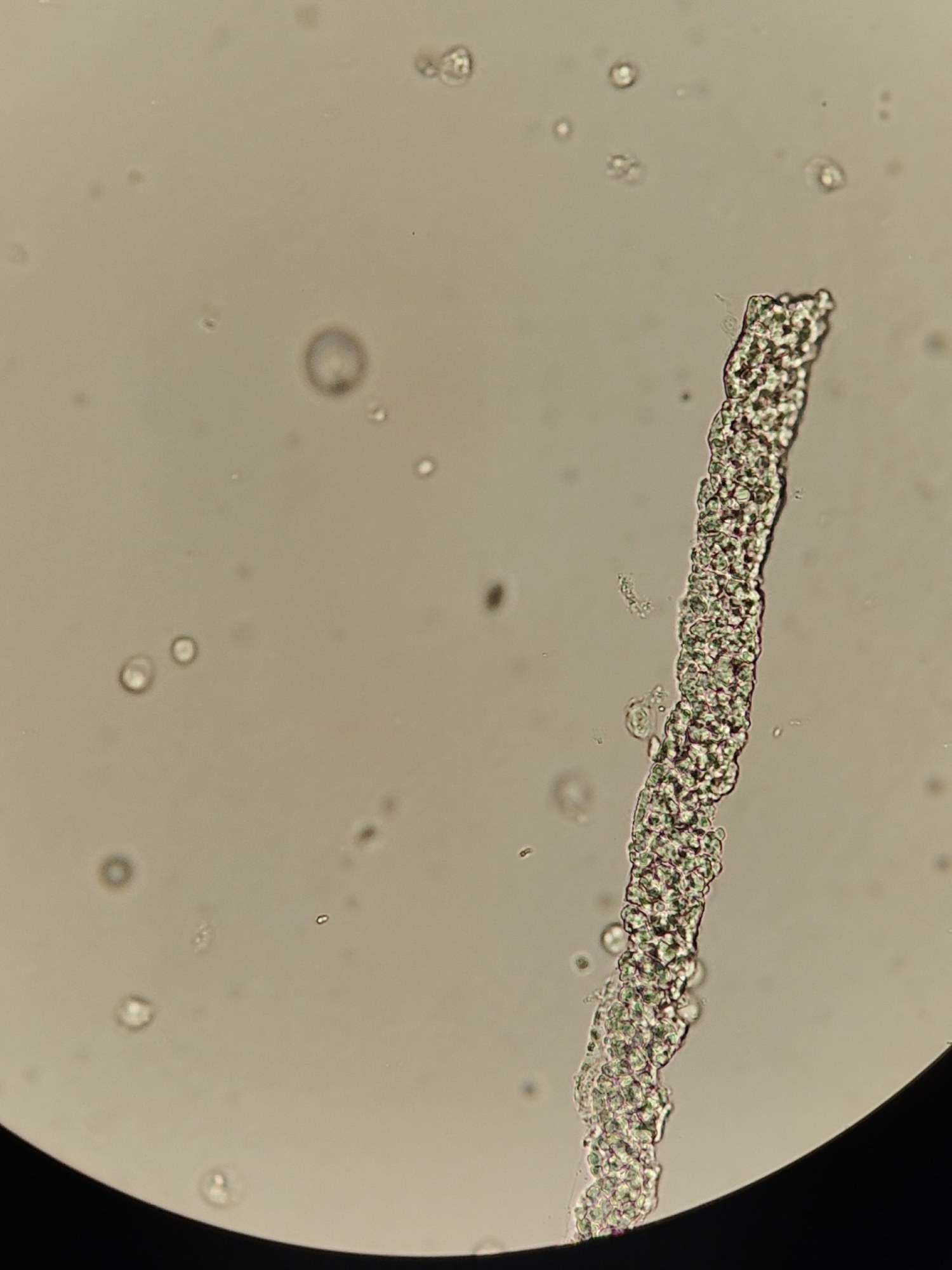
🔬 Urine Sediment Microscopy: Hyaline-Droplet Casts The photos show hyaline-droplet casts — structures formed from renal epithelial cells as a result of their breakdown during hyaline-droplet dystrophy.
Characteristics: Color: colorless or pale gray. Shape: cylindrical formations with an irregular contour. Internal inclusions in the form of flat droplets of the same or different sizes.
✅ Such findings may indicate impaired kidney function, including degenerative processes of the tubular epithelium.
🙏 Material and description kindly provided by biologist Anna Feditskaya, Luhansk.


