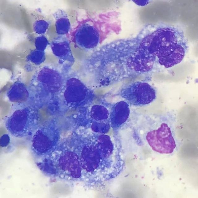
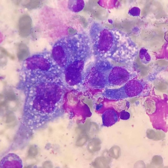
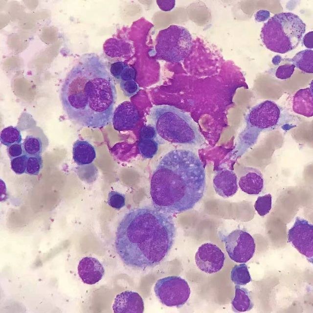
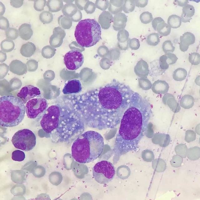
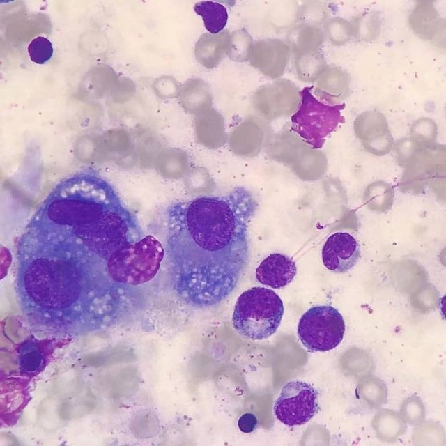
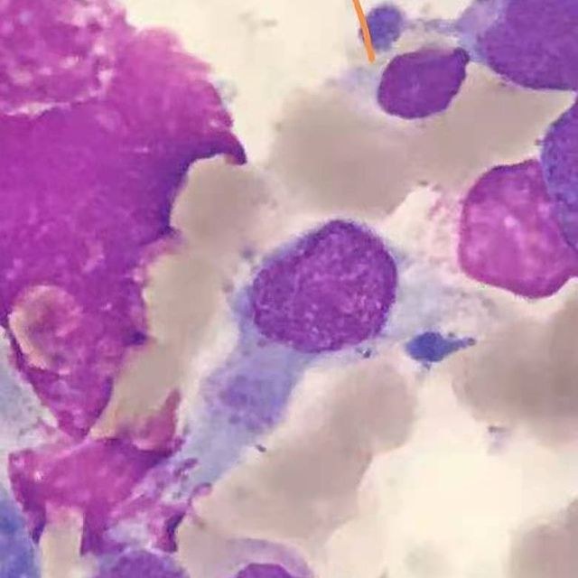
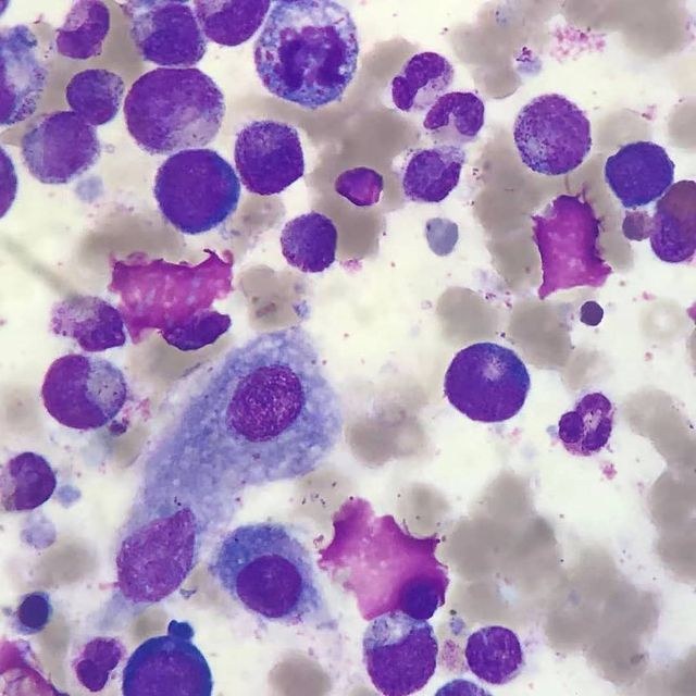
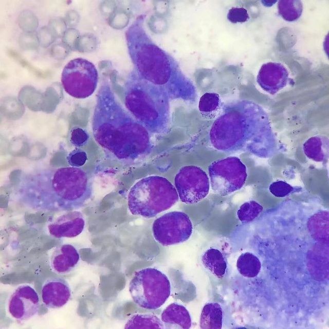
Langerhans Cell Histiocytosis. A rare disease, occurring in childhood at a frequency of 3-4 cases per 1 million pediatric population per year, even rarer in adults. The cause of the disease is unknown. A leading role is attributed to cytokines produced by T-lymphocytes and Langerhans cells in the lesions, which mediate the local accumulation of inflammatory infiltrates (eosinophils, macrophages, giant multinucleated cells). Clinical symptoms include bone lesions – skull vault, tibiae, ribs, pelvis, vertebrae, and mandible. Lymph nodes are involved in 30% of cases. Skin lesions in the form of dermatoses are observed in 30-40% of cases, less commonly lesions of the oral mucosa. Bone marrow involvement occurs in children with disseminated forms of the disease, accompanied by anemia, thrombocytopenia, and neutropenia. Morphological examination of bone marrow aspirate usually reveals normal cellular composition; isolated Langerhans cells may be found, but their diagnostic significance is low. Histological examination of bone marrow trephine biopsy is more informative. The diagnosis is confirmed by the detection of proliferating Langerhans cells expressing CD1a, langerin (CD207), S-100 protein, vimentin, CD68, and HLA-DR. Photo source inst:xiaoning.zhang Description source: Lugovskaya S. A., Pochtar M. E. Morphology of bone marrow cells in norm and pathology //Tver: Triada. – 2018.
Langerhans Cell Histiocytosis in Bone Marrow
Morphology of Langerhans cells in bone marrow aspirate, clinical manifestations, immunophenotype (CD1a, langerin), and diagnostic criteria for histiocytosis.