

🔬 Gram-negative diplococci, located both intracellularly and extracellularly, were found in a Gram-stained urethral smear from a male patient.
🙏 Image kindly provided by clinical laboratory diagnostics physician, Anna Ivanova, Moscow.
Microscopic image of a Gram-stained urethral smear showing intracellular and extracellular Gram-negative diplococci (gonococci) and neutrophils.
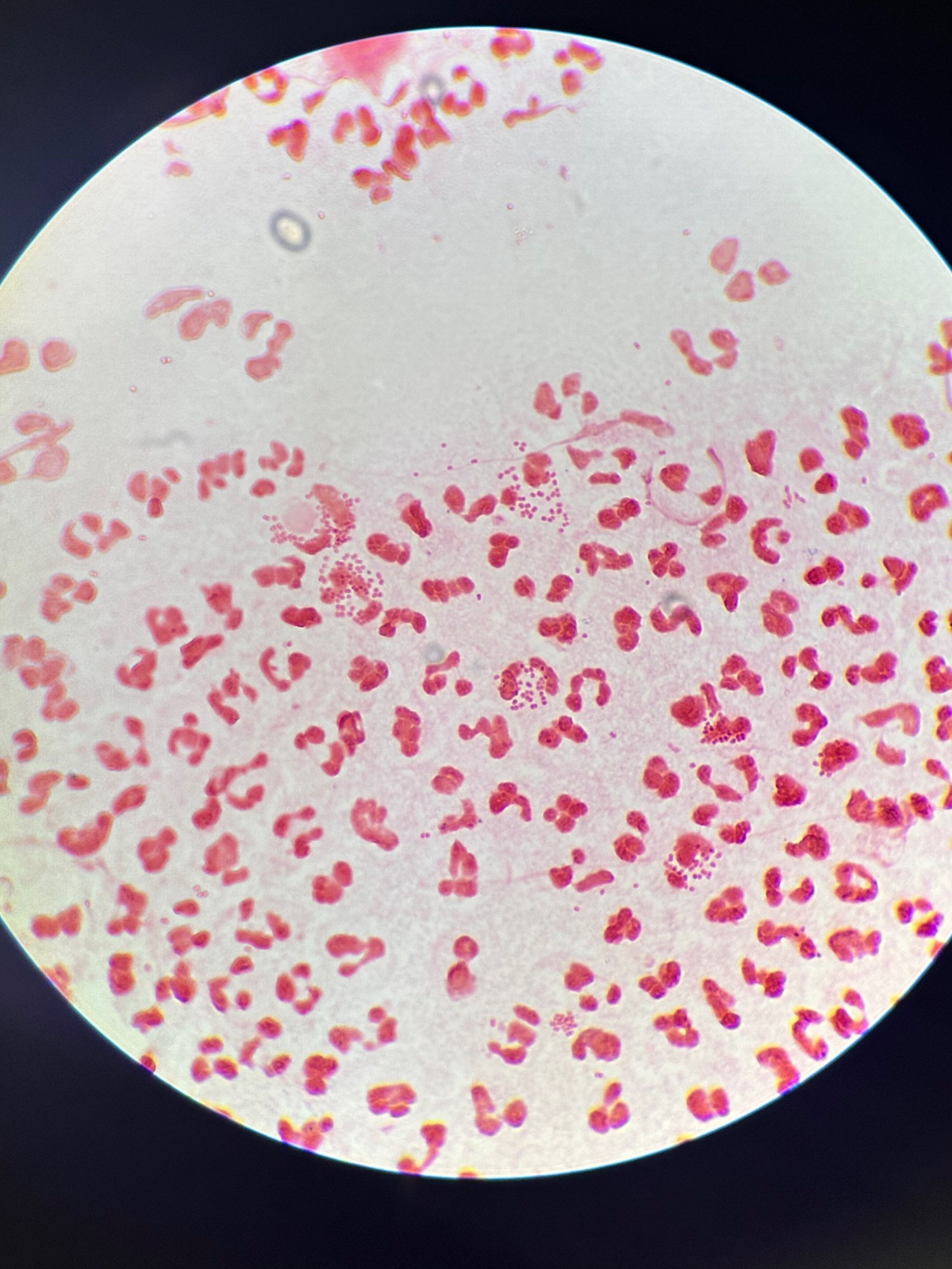
🔬 Gram-negative diplococci, located both intracellularly and extracellularly, were found in a Gram-stained urethral smear from a male patient.
🙏 Image kindly provided by clinical laboratory diagnostics physician, Anna Ivanova, Moscow.

Microscopic view of a male urethral smear in gonorrhea: pronounced leukocytosis and phagocytosis of kidney-shaped diplococci (Neisseria gonorrhoeae) by neutrophils.

Microscopic findings of acute gonorrhea: detection of intracellular and extracellular Neisseria gonorrhoeae in a male urethral smear stained with methylene blue.

Microscopic detection of Neisseria gonorrhoeae in urogenital smears: features of methylene blue and Gram staining, and diplococci phagocytosis.