

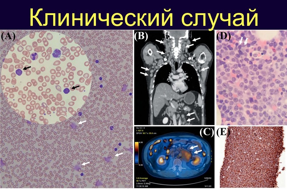
Chronic lymphocytic leukemia (CLL). A 54-year-old male entrepreneur. Since 2003 – generalized lymphadenopathy, occasionally received chlorambucil from a private doctor. In 2017, he presented with:
- increasing lymphadenopathy (neck, axillary, and inguinal regions)
- night sweats for 6 months
Liver and spleen were not palpable.
✅ Complete Blood Count at presentation (2017)
- Hemoglobin: 13.4 g/dL (within normal limits)
- Leukocytes: 19.4 × 10⁹/L
- Neutrophils: 3.44 × 10⁹/L
- Lymphocytes: 15.2 × 10⁹/L (marked lymphocytosis)
- Platelets: 139 × 10⁹/L (moderate decrease)
✅ Peripheral Blood Smear (Fig. A)
- Numerous small mature-appearing lymphocytes
- dense, “clumped” chromatin
- high nuclear-to-cytoplasmic ratio
- scanty, narrow cytoplasm
- Readily identifiable “smudge” cells – Gumprecht shadows
Morphological picture is typical for chronic lymphocytic leukemia.
✅ Imaging Positron emission tomography combined with computed tomography showed:
- enlarged lymph nodes up to 3 cm
- localization: neck, axillary, and inguinal regions
- moderate metabolism (eumetabolic lymph nodes) – no signs of aggressive transformation at baseline (Fig. B).
✅ Diagnosis Confirmation Bone marrow examination was performed:
- predominance of small lymphocytes
- immunophenotype characteristic of chronic lymphocytic leukemia according to flow cytometry data:
- co-expression of CD5 and B-cell markers
- CD23 positive
- weak expression of surface immunoglobulin, CD20, CD79b
- FMC7 negative or weak
Cytogenetics (fluorescence in situ hybridization):
- no 11q and 17p deletions
- no trisomy 12
- no 13q deletion (i.e., no classic “favorable” variant)
Molecular markers:
- immunoglobulin heavy chain gene (IGHV) – mutated variant, which is associated with a more favorable prognosis.
Conclusion: chronic lymphocytic leukemia without unfavorable cytogenetic markers, with mutated IGHV.
✅ Initial Therapy and Response In 2017, the patient received 6 courses of immunochemotherapy with fludarabine–cyclophosphamide–rituximab. Complications:
- pneumonia
- episode of cytomegalovirus antigenaemia (due to immunosuppression)
Upon completion of treatment:
- bone marrow retained lymphocytic infiltration of about 20%
- cells lost CD20 expression (expected after rituximab therapy)
- peripheral lymphocytosis was absent
- lymph nodes were not palpable, no general symptoms
- according to positron emission tomography combined with computed tomography: significant reduction in size and metabolism of lymph nodes (to ~1 cm, eumetabolic).
The patient was prescribed long-term treatment with the Bruton’s tyrosine kinase inhibitor ibrutinib.
✅ Richter’s Syndrome Transformation (2023) Several years later, during therapy, the following developed:
- progressive lymphocytosis
- sharp increase in lactate dehydrogenase activity to 1200 U/L (normal 150–300 U/L)
- appearance of large atypical lymphocytes in the blood
Positron emission tomography combined with computed tomography:
- enlarged, hypermetabolic para-aortic lymph nodes (Fig. C)
Bone marrow and lymph node biopsies (under computed tomography guidance):
- in the bone marrow – sheets of large atypical lymphoid cells (Fig. D)
- immunohistochemistry: large cells express CD20 (Fig. E)
- picture consistent with transformation of chronic lymphocytic leukemia into aggressive large B-cell lymphoma (Richter’s syndrome).
Next-generation sequencing:
- two TP53 gene mutations detected in the bone marrow – molecular explanation for aggressive course and resistance.
✅ What is important for a laboratory hematologist in this case ☑️ Morphology
- Lymphocytosis of small mature-appearing lymphocytes with Botkin-Gumprecht shadows, with preserved erythrocytes and platelets in an adult patient – always a reason to consider chronic lymphocytic leukemia and refer for flow cytometry.
☑️ Immunophenotype and Genetics
- Diagnosis confirmation – by characteristic B-cell immunophenotype with co-expression of CD5 and CD23 and weak expression of surface immunoglobulin.
- Mandatory search for 17p and 11q deletions, trisomy 12, 13q deletion; evaluation of IGHV status and TP53 mutations – this determines prognosis and choice of targeted therapy.
☑️ Dynamic Observation
- Rapid increase in lymphocytosis, sudden elevation of lactate dehydrogenase activity, appearance of large blast-like cells, and localized increase in lymphadenopathy – laboratory “red flags” for possible Richter’s syndrome transformation.
- At these stages, a close link between morphology, flow cytometry, histology, and molecular diagnostics is crucial.
☑️ Role of Imaging
- Positron emission tomography combined with computed tomography at baseline allows assessment of lymphoid tissue volume.
- In case of suspected transformation – search for foci with high metabolic index for targeted biopsy.
Such a case well illustrates the journey of chronic lymphocytic leukemia: from “benign” lymphocytosis with a typical blood smear to targeted therapy and, ultimately, to Richter’s syndrome, where it is critical for the laboratory service not to miss the moment of change in tumor morphology and biological properties. Source: Anskar Y.H. Leung - Haematology and the Asian Patient 51 Clinical Cases - 2024


