

The future of hematological diagnostics: artificial intelligence in bone marrow analysis 🧬🔬
Traditional microscopy is the “gold standard” for diagnosis, but it is not immune to subjectivity and requires enormous time. The International Journal of Laboratory Hematology published the results of a study on the Scopio X100 HT digital morphology system. Scientists from Rome investigated how effectively algorithms cope with the analysis of bone marrow aspirates in onco-hematological pathologies.
📊 Study essence Doctors analyzed 334 bone marrow samples from patients with various diagnoses: from myelodysplastic neoplasms to acute leukemias. The system scans the smear, creates an ultra-high-resolution digital image, and automatically categorizes cells. The obtained data were compared with manual counts performed by expert hematologists.
⚖️ Key results:
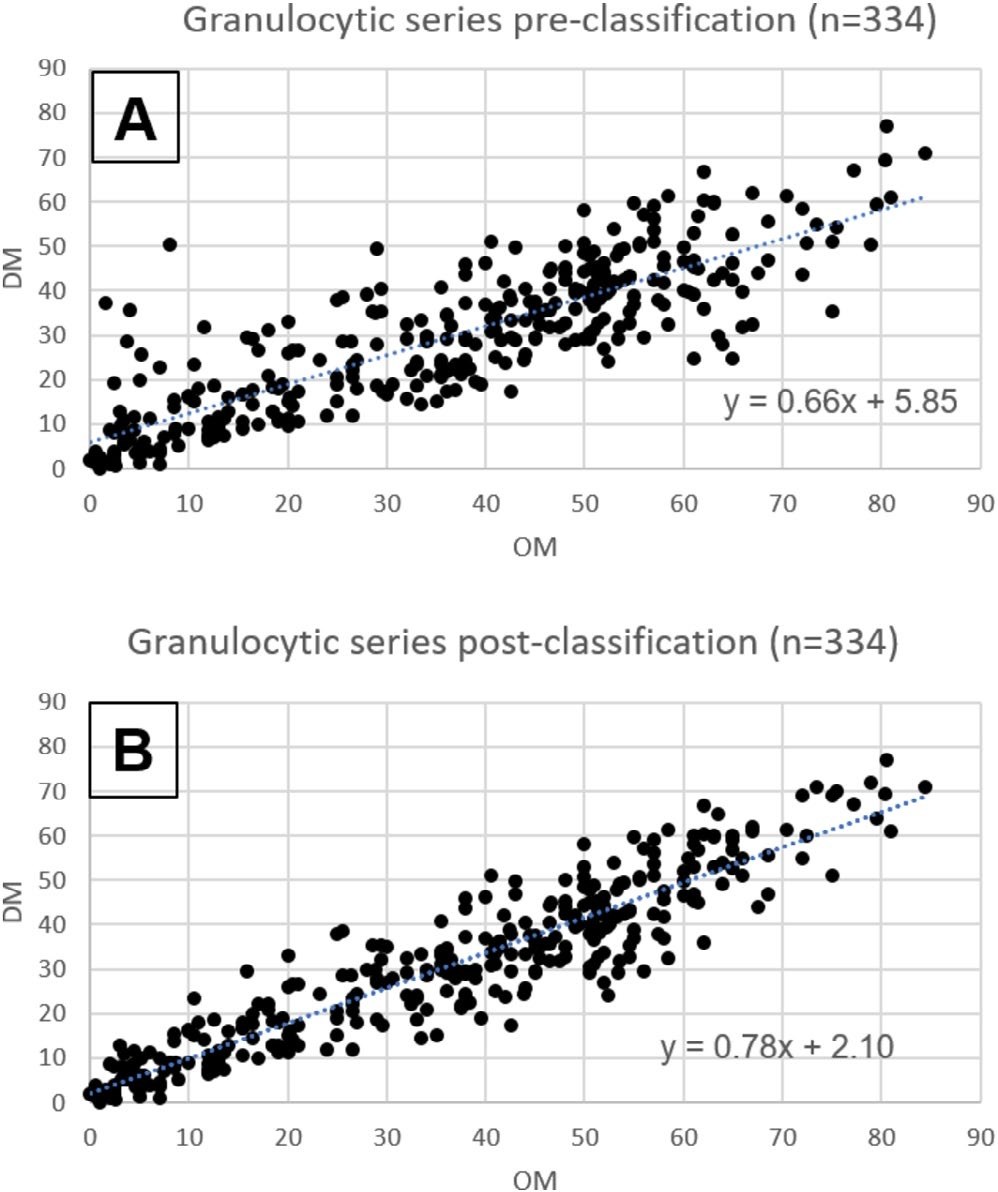
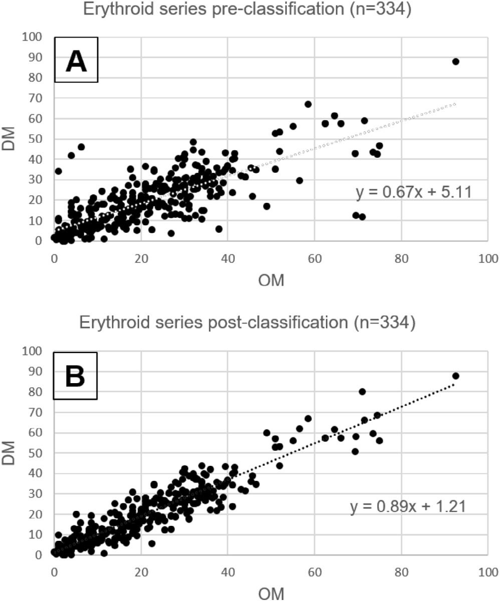
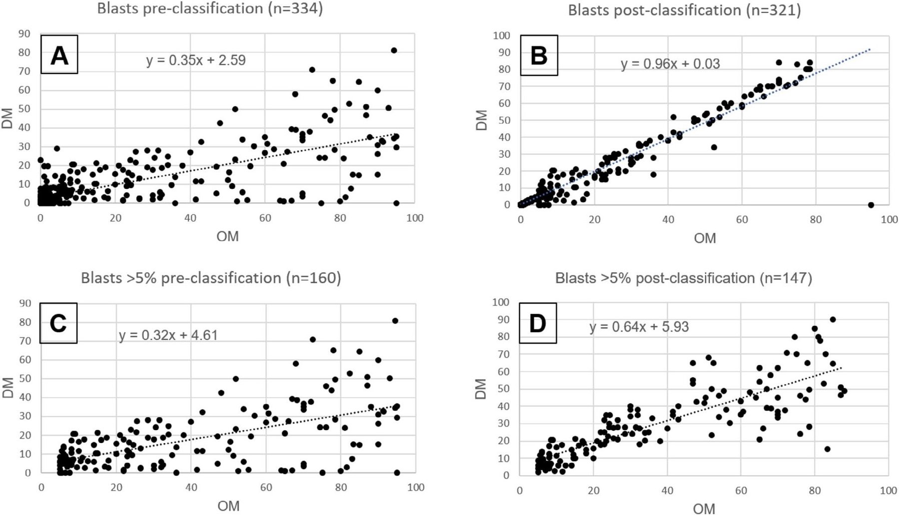
🔹 Granulocytic lineage: The highest correlation with the manual method was shown. After physician review, accuracy reached almost maximum (r = 0.93). 🔹 Erythroblasts: Artificial intelligence successfully recognized red blood cells, even in the presence of dyserythropoiesis signs (morphological anomalies). 🔹 Blast cells: In samples with blast content less than 5%, the system worked flawlessly. In diagnostically complex cases with high blastemia (>5%), the accuracy of automatic determination was lower but significantly increased after the physician’s “post-classification” stage. 🔹 Megakaryocytes: The digital system proved extremely useful for finding rare giant cells. Where a physician’s eye might miss them during normal viewing, full-field scanning found single elements.
🚩 Difficulties and limitations The system is not yet fully autonomous. Pronounced hypercellularity of smears (very high cell density) and atypical lymphoid populations still require close attention from a specialist. Also, morphological differentiation of monoblasts and promonocytes remains a challenging task for current algorithms.
🎓 Conclusion for clinicians Digital morphology does not replace the physician but radically changes the workflow. It allows: ✅ Standardizing myelogram counting. ✅ Creating digital archives for dynamic patient monitoring. ✅ Conducting remote consultations (telemedicine) without sending slides.
Artificial intelligence is becoming a reliable assistant, minimizing the human factor and accelerating diagnosis in complex clinical cases. 🏥🤖
Source for citation: Zini G, Chiusolo P, Marra J, Rossi E, Bellesi S, Pelliccioni N, d’Onofrio G. Assessment of the Performance of Siemens Scopio Digital Morphology on Bone Marrow Aspirates in Onco-Hematology. International Journal of Laboratory Hematology, 2026; 0:1–8. DOWNLOAD PDF


