

Clinical case on cytochemistry from 🇺🇿 Uzbekistan
❗ Patient, 62 years old. Complaints upon admission: frequent headaches, dizziness, elevated blood pressure, skin itching, skin redness, weakness, and fatigue.
✅ Preliminary diagnosis: polycythemia vera.
🩸 Hemoglobin 189 g/L. Erythrocytes 6.8 million/mL. Leukocytes 5.6 thousand/µL. Thrombocytosis. ESR 3 mm/hour.
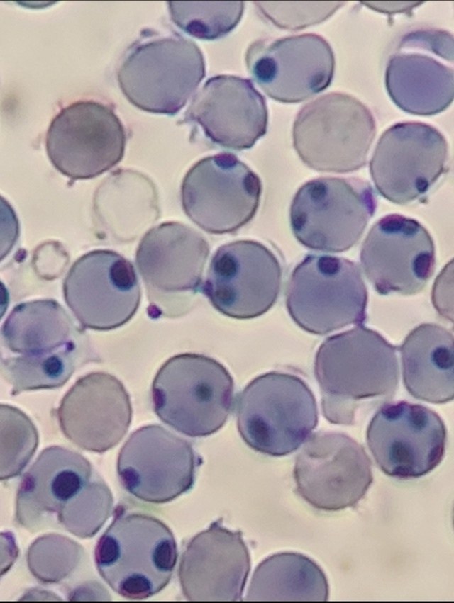
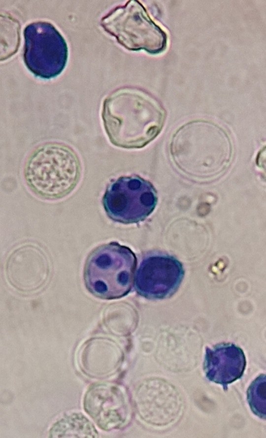
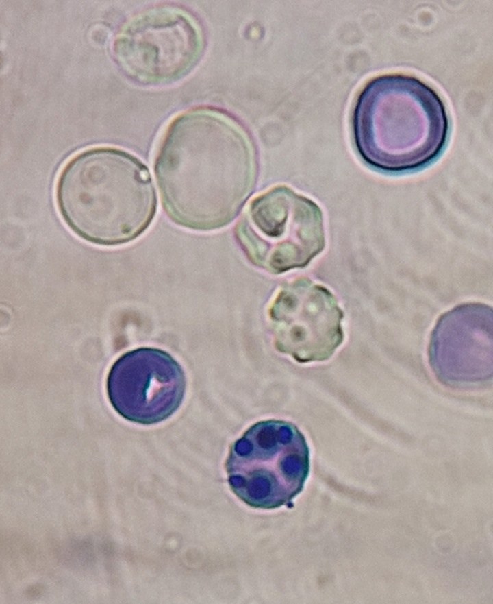
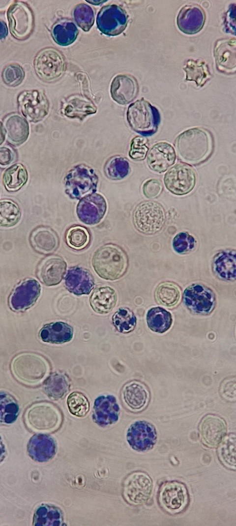
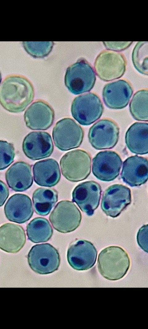
🎨 Erythrocyte cytochemistry: sodium nitrite treatment, performed to convert hemoglobin to methemoglobin, showed an increase in the number of erythrocytes with methemoglobin (images below).
❓What additional tests are needed to confirm the diagnosis?
🙏 Clinical case kindly provided by Zebiniso Kholmuradova, morphologist, Uzbekistan, Kashkadarya Multidisciplinary Children’s Medical Center.
📖 Reference data: Method for erythrocyte examination with sodium nitrite for methemoglobin detection Determination of the ability of erythrocytes to accumulate methemoglobin by cytochemical conversion of hemoglobin to methemoglobin using sodium nitrite.
Equipment and reagents: Sodium nitrite (NaNO₂) solution — 1% solution. Dry glass slides. Fixative — a 1:1 mixture of alcohol and acetone can be used. Stain — optional, usually hematoxylin-eosin or another staining agent for cell differentiation. Microscope — light microscope with up to 1000x magnification. Procedure: Blood collection:
Use fresh capillary or venous patient blood, anticoagulated (e.g., with EDTA). Smear preparation:
Apply a drop of blood to a clean slide and make a smear using a standard method. Allow the smear to air dry.
Fixation: Fix the smear by immersing it for 5–10 minutes in fixative (1:1 alcohol and acetone mixture). Allow the smear to air dry. Sodium nitrite treatment:
Immerse the smear in a 1% sodium nitrite solution for 10–15 minutes. Sodium nitrite oxidizes hemoglobin to methemoglobin, giving erythrocytes a specific staining that can be visualized microscopically.
Staining: After sodium nitrite treatment, rinse the smear with distilled water to remove excess reagent. Stain the smear with a standard stain (e.g., hematoxylin-eosin) to improve contrast and cell identification.
Microscopy: Dry the smear and examine it under a light microscope at 400–1000x magnification. Look for erythrocytes containing methemoglobin. Such cells may differ in color (darker areas) due to the presence of methemoglobin, which forms after sodium nitrite treatment.
Interpretation of results: Positive result: An increased number of erythrocytes containing methemoglobin is observed. This indicates a potential tendency of erythrocytes to oxidize hemoglobin to methemoglobin, which may be associated with cell membrane characteristics or enzymatic disorders. Negative result: Erythrocytes remain unstained by methemoglobin or only an insignificant amount is present. This is considered normal and indicates no increased tendency to accumulate methemoglobin.
Note: This method is not an indicator of the patient’s blood methemoglobin level. It is used to detect the resistance of erythrocytes to oxidative damage and the ability of cells to accumulate methemoglobin under artificial exposure.
Important: Since the method is not standard, its results should be interpreted with caution and considered in conjunction with clinical and laboratory data.


