

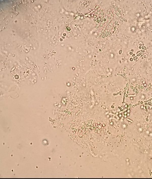
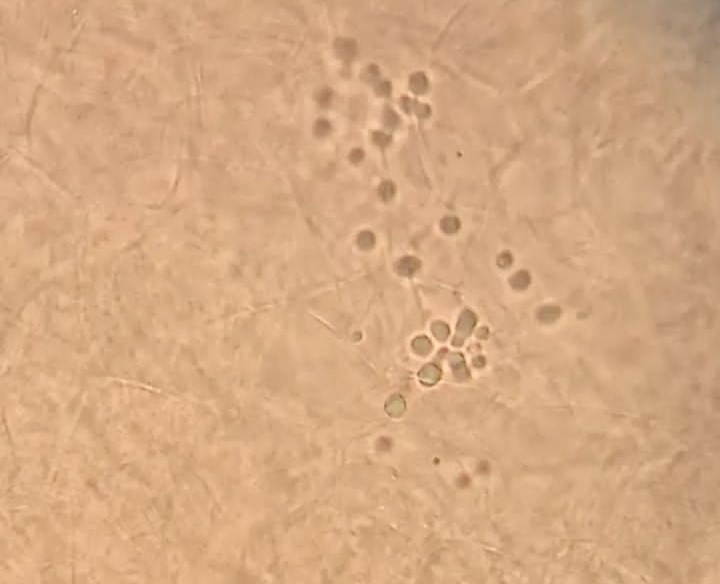
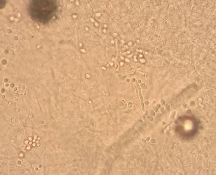
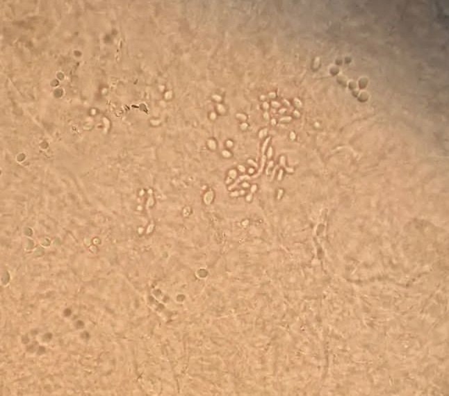
🔬 Blastospores (budding cells) and pseudomycelium of Candida fungi. Fingernail plate (photo 1) and interdigital spaces of the feet (photos 2,3,4), KOH 20%, magnification x400+zoom.
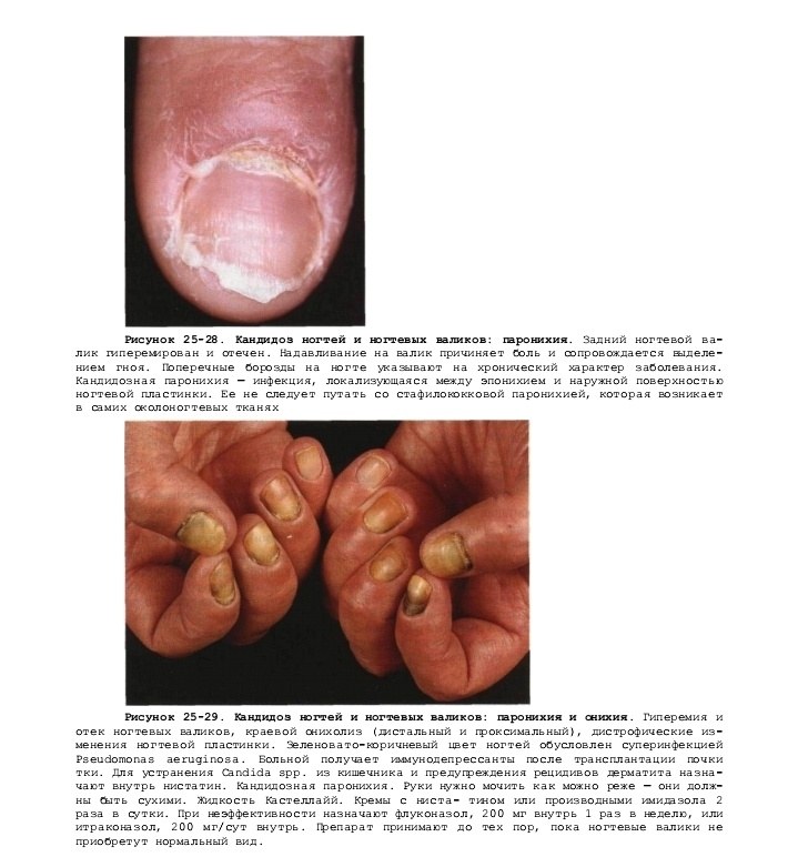
✅ In candidal onychomycosis, paronychia (inflammation of the proximal nail fold) precedes onychomycosis. First, the nail fold reddens, becomes shiny, thickens and swells, changing its shape, which causes the cuticle to separate from the nail plate and break down. Inflammation of the nail fold may be accompanied by pain. The course of paronychia is undulating; the nail is gradually involved.
🙏 Material kindly provided by medical laboratory technician Dmitry Emelyanov.


