

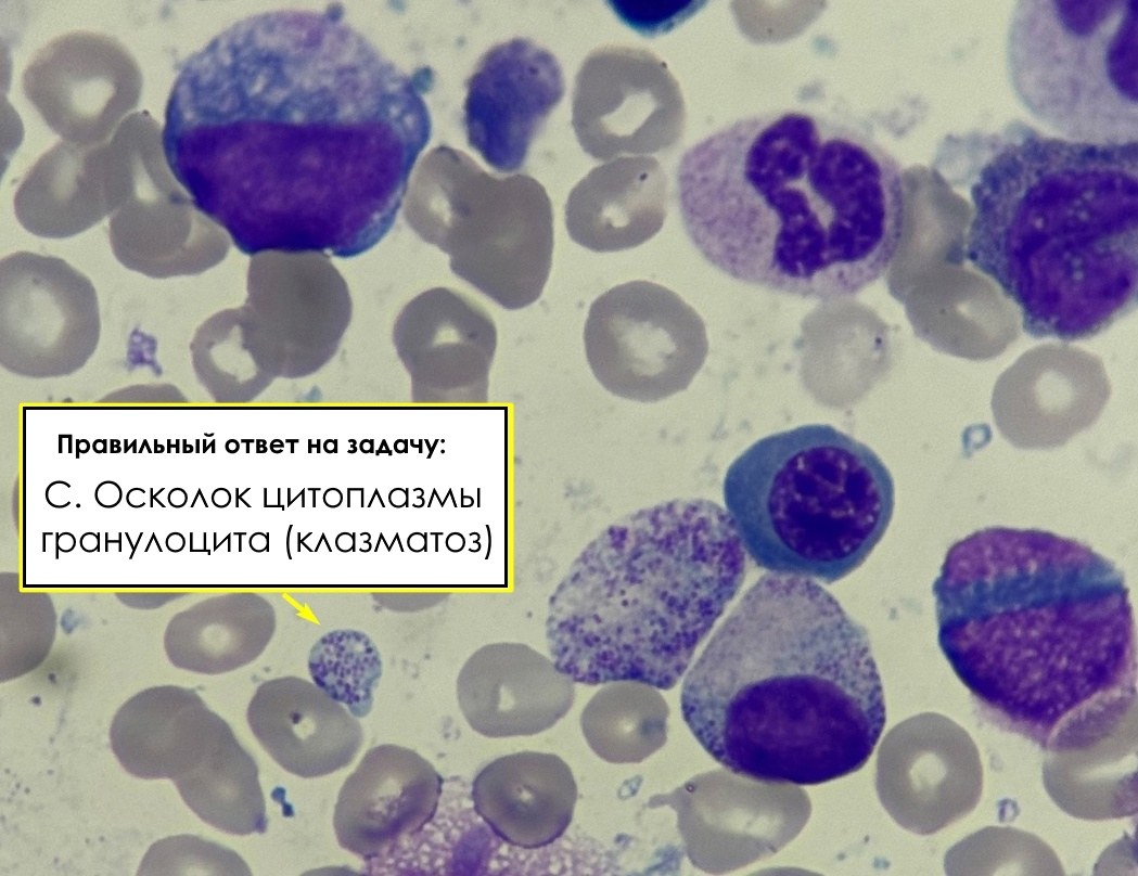
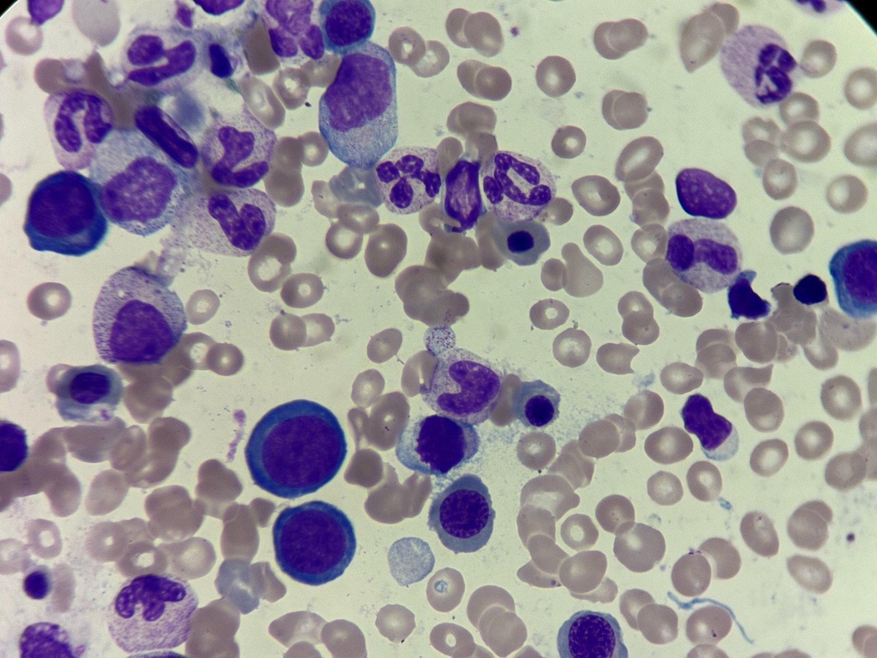
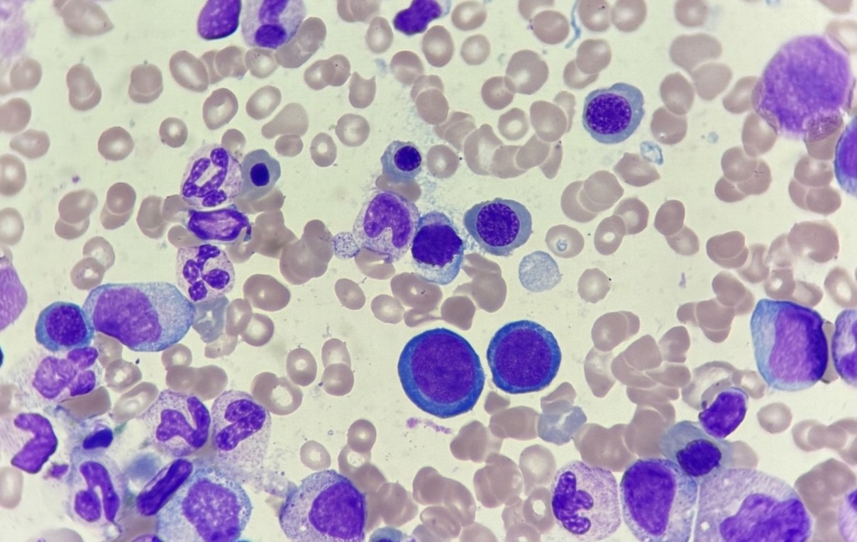
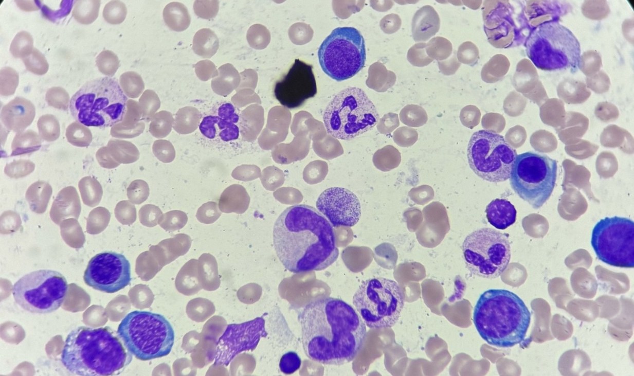
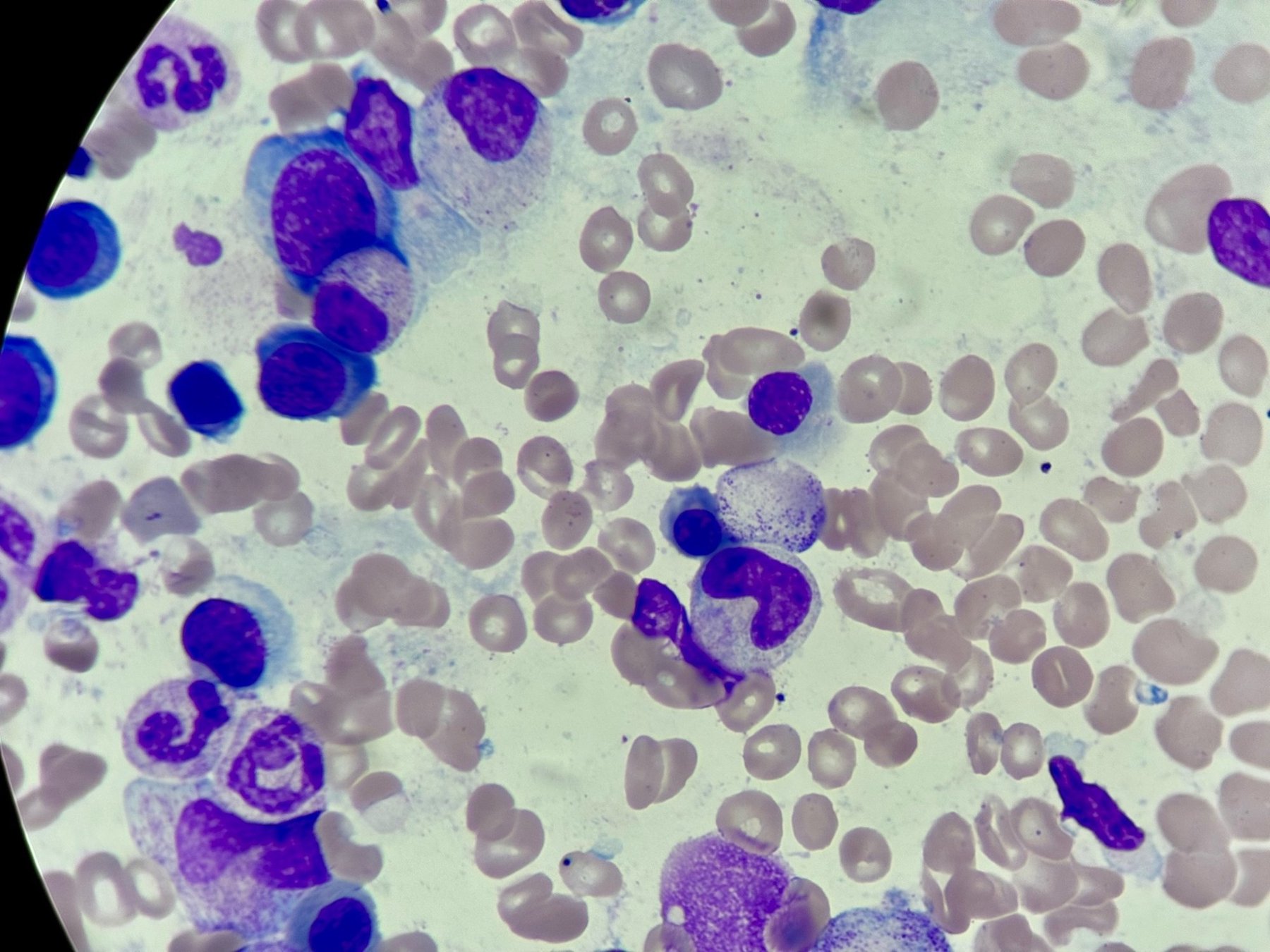
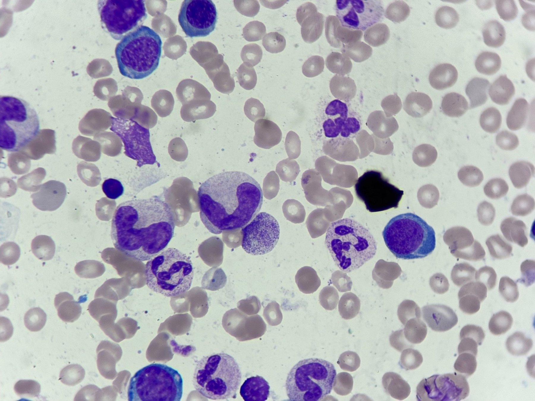
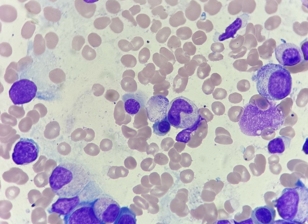
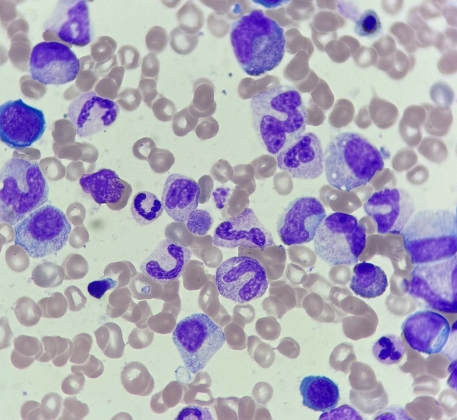
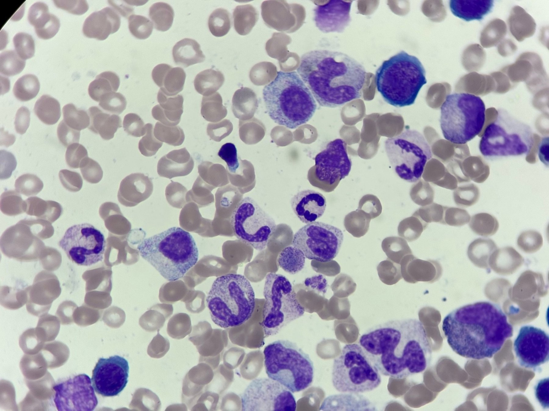
Correct answer to the problem from the closed club of laboratory hematologists. C. Granulocyte cytoplasmic fragment (clasmacytosis) In the image, in the lower left part of the field, there is an isolated rounded fragment with basophilic cytoplasm, devoid of a nucleus. To its right, a large cytoplasmic fragment with characteristic granulation is clearly visible, which allows interpreting the finding as a fragment of the cytoplasm of a mature granulocyte — a manifestation of clasmacytosis.
☝ The phenomenon of clasmacytosis is the fragmentation of a cell’s cytoplasm, in which fragments (cytoplasmic bodies) separate from the main cell body. This can be a consequence of active phagocytosis, toxic effects, or cytolysis against a background of severe conditions.
📚 Important to know: clasmacytosis is not only granulocytic.
This phenomenon is also characteristic of plasma cells and lymphocytes:
🔸 In plasma cells, rounded cytoplasmic fragments can separate, especially during their increased activity, for example, in plasmacytosis or myeloma.
🔹 In lymphocytes, such fragments are called lymphoglandular bodies — rounded, homogeneous pieces of cytoplasm without a nucleus, often found in smears of lymph node and bone marrow aspirates.
🔻 Why not other options? A. Erythrocyte with basophilic stippling ✘ The contour and shape of the object do not correspond to an erythrocyte; uniform cytoplasm and typical fine stippling are absent.
B. Macroplatelet ✘ Macroplatelets appear as large homogeneous fragments, often with granules and without an adjacent cytoplasmic fragment. Here — there is a clear granulocytic context.
D. Malarial plasmodium ✘ The parasite has a different shape and structure.
The patient has a reduced folic acid level, with signs of dyserythropoiesis in the myelogram (other aspirate images in the carousel).
🏆 Congratulations to the winners! They receive a private invitation to the club of selected specialists:
- Irina Korolkova - subscriber of VK donut Leukformula, already has access to the closed club;
- Sergey Ermakov - invitation to the closed club sent;
- Asya Nikolaeva - private messages are closed. Please write to the instructor to receive an invitation;
- Elena Ionova - invitation sent to private messages;
- Maria Eliseeva-Shatskikh - invitation sent to private messages.
🙏 Smear photos for the task were kindly provided by Kristina Yurievna, laboratory physician, BU “RKB”, Cheboksary


